From Blade To Electrode
Oct 02, 2026
The operating theater once defined gynecology. Blades, clamps, suture reels, suction canisters, the hum of an anesthetic machine - this was the visual language of fibroid treatment, and with it came admission days, blood-transfusion anxiety, adhesion risk, and the slow recovery that steals a month of a woman's working life. For younger patients, the unspoken dread was fertility: a myomectomy could scar the endometrial cavity; a hysterectomy closed the question entirely. Older patients with hypertension, diabetes, or cardiac comorbidities were too often told they were poor surgical candidates and left to manage menorrhagia with tranexamic acid and resignation. Layered on top was a quieter form of over-treatment: small intramural fibroids causing only mild pressure were sometimes routed straight to hysterectomy simply because no precise middle ground existed in the local toolkit. The gap was not a lack of courage; it was a lack of a scalpel-free option with predictable boundaries.
Radiofrequency puncture needles fill that gap by abandoning mechanical cutting altogether. There is no blade edge parting tissue; instead, high-frequency current turns tissue water into a resistive heater. Ions oscillate, friction accumulates, and temperature climbs. Once the fibroid core sustains above 60°C, smooth-muscle cells lose membrane integrity, collagen stroma coagulates, and microvessels occlude. As heating proceeds, the local pH falls, hormone receptors are denatured, and the nodule's ability to respond to estrogen wanes. The generator is not blind to this: as tissue dehydrates, impedance rises, and a well-designed unit rolls off power to prevent charring and gas cavitation. What the surgeon feels is almost nothing; what the fibroid experiences is a controlled, lethal fever. The uterus, kept cool at its margins, retains its contour and, in most cases, its future potential.
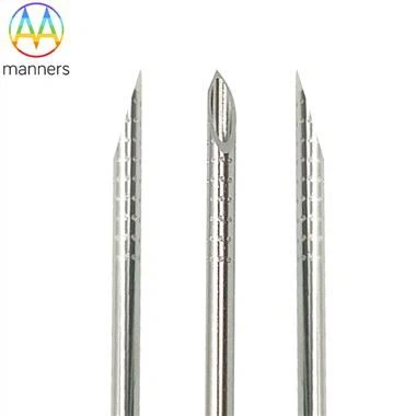
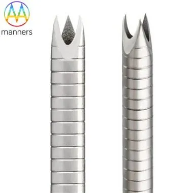
Classification here is less about brand names and more about how energy returns and how the shaft behaves. Fixed-power needles deliver a set wattage and rely on the operator's timing; impedance-controlled needles sense tissue resistance and modulate automatically; temperature-sensing needles embed a thermocouple at the tip to cap the core temperature. Expandable multi-array electrodes transform a single puncture into a three- or four-prong fan, widening the ablation ellipse without additional skin entries. By gauge, 8G to 10G shafts are stiff and suited to large deep lesions, 14G to 18G form the interventional middle ground, and 21G to 26G offer fine, low-pain access for superficial or cervical routes. Manufacturing distinctions matter as much as clinical ones: CNC-turned shafts hold concentricity; swaged hubs prevent leakage at the connector; laser-etched depth rings give the operator millimeter confidence; electropolishing lowers the Ra value so the needle does not drag thrombus back up the track; and validated EO sterilization turns a metal tube into a clinical-grade single-use device.
The operative choreography rewards patience over aggression. Pre-operatively, a CBC, coagulation profile, and a negative pregnancy test are non-negotiable; pelvic ultrasound defines the safety envelope, and MRI is reserved for ambiguous subserosal or cornual lesions. In the room, the gel is warmed to reduce artifact, the bladder strategy is decided in advance - filled to window, emptied to retreat. Entry routes are chosen by anatomy: transabdominal for anterior and posterior intramural masses, transvaginal for cervical and submucosal lesions. Energy begins low; the sonographer calls out bubble formation like a co-pilot. If pain spikes or the hyperechoic zone creeps toward the serosa, power drops or the needle repositions. A saline window is injected when bowel or bladder lies within a centimeter of the thermal margin. The procedure ends not with a suture but with a scan: is the lesion now uniformly hyperechoic, is perfusion gone at the core?
Those who cross-train from thyroid and hepatic ablation bring a shared intuition: respect the heat sink. Vessels carry heat away, so perivascular tissue needs a touch more energy; bowel and bladder have no such tolerance and must be shielded. Hydrodissection, once considered an extra step, has become standard wisdom in experienced hands. The most common disappointment is not failure but expectation - patients who expect the nodule to disappear on the two-week scan. Clinicians now preface every consent with a timeline: symptoms ease first, volume shrinks over three to six months, the imaging shadow lingers longest. One multi-center observation is consistent: centers that treat ablation as chronic-disease management, not a one-minute fix, report the steadiest satisfaction scores.
Elevating the perspective, the electrode becomes a scalpel made of temperature. The surgeon's skill is no longer measured in the force of the cut but in the geography of heat - where it goes, where it stops, what it spares. This is a gentler kind of mastery, and arguably a more modern one, suited to an era where patients ask not "what will you remove?" but "what will you preserve?"
Recommendations for the coming cycle are pragmatic. Form joint gynecology–radiology–anesthesia ablation teams rather than lone-wolf operators. Procure needles with documented thermal profiles and labeled exposed-tip lengths. Regulators should require ablation-zone validation in tissue-mimicking phantom, not merely a sharpness test. And residencies should teach "thermal literacy" alongside suturing, because the future of fibroid care will increasingly be written in degrees Celsius.