Needles That Burn, Not Cut
Oct 02, 2026
For decades, the clinical conversation around symptomatic uterine fibroids has been framed by a grim binary: tolerate the heavy menstrual bleeding, the dragging pelvic pressure, the iron-deficiency anemia that leaves a woman short of breath climbing stairs, or submit to a scalpel that may take the uterus away entirely. Myomectomy offers organ preservation but demands laparotomy or laparoscopy, general anesthesia, a hospital stay, and the lingering risk of adhesion formation and fibroid recurrence. Hysterectomy, meanwhile, settles the mass permanently but erases fertility and carries its own psychological weight that is too often dismissed in the operating room corridor. Between these two extremes, a generation of patients asked a deceptively simple question - could the fibroid simply be switched off without removing the womb? The frustration was not only medical but logistical: young women postponing childbearing, perimenopausal patients deemed "too anemic to operate," and clinicians wary of offering minimally invasive options that lacked reproducible precision. Add to this the older problem of blind puncture: without real-time imaging, a needle could skim past the lesion, graze the endometrium, overheat the serosa, or leave a viable rim of fibroid tissue that would quietly regrow. Needle rigidity varied between batches, tip bevels were inconsistent, and electrodes without proper insulation burned the track on the way in. These were not minor annoyances; they were the reasons many centers hesitated to adopt thermal therapy at all.
The physics that resolves this tension is deceptively quiet. A radiofrequency generator emits alternating current in the 375 to 500 kHz range. Inside tissue, ions cannot follow the field instantaneously; they oscillate, collide, and generate frictional heat. As the local temperature crosses roughly 60°C, intracellular proteins begin to denature; between 65 and 100°C, coagulative necrosis becomes irreversible. Guided by B-ultrasound, the radiofrequency puncture needle is advanced to the fibroid core, and the thermal field radiates outward in a predictable ellipsoid. Water leaves the cells, the stroma dehydrates, small feeding vessels thrombose, local nerve endings are damaged, tissue pH drops, and estrogen/progesterone receptors in the fibroid are destroyed. What remains is a biologically inert nodule that the body's phagocytic immune response gradually resorbs or shrinks. The uterus keeps its architecture; only the lesion loses its drive to grow. This is not vaporization and not excision - it is thermal inactivation, a shift from removing tissue to retiring its function.
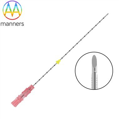
Yet "RF needle" is never a single object. The monopolar single-tip needle, tethered to a dispersive grounding pad, delivers a focused thermal lesion and remains a workhorse for solitary intramural fibroids. The monopolar multi-claw electrode expands inside the nodule, opening two to four prongs to sweep a larger ellipsoidal death zone - indispensable for 4 to 6 cm lesions. Bipolar needles route current between two active tips on the same shaft, dispensing with the patient pad and suiting narrow-field, transvaginal work. Straight rigid stainless-steel needles serve transabdominal approaches where the shaft must resist deflection through the abdominal wall; flexible curved electrodes navigate the natural cervical route toward submucosal masses. Beneath these clinical forms sits the manufacturing layer that rarely makes it into marketing copy: 316LVM stainless tubing, necked-down distal segments produced by rotary swaging, precision tip grinding for a clean entry bevel, laser cutting for side-window configurations, laser marking for centimeter depth rings, electropolishing to drop surface roughness and reduce thrombus adherence, and finally validated sterilization - EO or gamma - in a single-use pouch. Gauge spans 8G for robust large-bore access down to 26G for fine, low-trauma tracks, with OEM programs translating a clinician's 2D drawing or 3D sample into a reproducible batch.
In practice, the procedure begins long before the needle touches skin. A complete blood count and coagulation panel set the anemia and bleeding-risk baseline; a pelvic ultrasound - ideally supplemented by MRI - maps fibroid number, depth, distance from serosa, endometrium, bladder, and rectosigmoid. The operator chooses a trajectory that threads through relatively avascular myometrium. Local anesthesia is administered, the bladder is partially filled to act as an acoustic window or deliberately emptied to drop the dome away from an anterior fibroid. Power is ramped gradually rather than switched to maximum; the sonographer watches for the first faint hyperechoic microbubbles as proof the field is forming, then modulates energy to avoid cavitation. For lesions over 5 cm, the needle is withdrawn in steps - a "moving ablation" fan - so overlapping ellipses cover the whole mass. A saline hydrodissection pocket is sometimes created to push bowel away from the thermal margin. Post-procedure, contrast-enhanced ultrasound or early MRI quantifies the non-perfused volume, the truest marker of success.
Clinicians who have performed hundreds of these cases converge on an almost Zen-like discipline. The best ablations look uneventful: a quiet room, a steady hand, a needle that barely seems to move. One reported case of an 11 cm fibroid used multipoint moving ablation over roughly fifty minutes, leaving nothing but a 1 mm skin mark. Experienced operators repeat a mantra - gas is information. A fine bubble cloud means the field is alive; sudden boiling means you have overshot. They also resist the temptation to promise a vanished mass on the next scan; coagulated fibroid tissue can linger as a shadow on ultrasound for months even as symptoms improve dramatically. The lesson hard-won across centers is that the needle accounts for perhaps thirty percent of the outcome; the remaining seventy is ultrasound literacy, trajectory restraint, and the judgment to decline ablation when a subserosal pedunculated fibroid sits too close to the bowel.
There is a quiet philosophical shift embedded in all of this. Surgery has long equated healing with removal - take out the diseased part, and the body is made whole. Radiofrequency puncture needles invert that logic: the organ stays, the lesion is silenced. The wound is not a scar measured in centimeters but a track measured in millimeters. For women who have delayed care for fear of losing their uterus, that reframing is as therapeutic as the heat itself.
Looking forward, the recommendation is not "buy more generators" but "build teams that can see." Hospitals should credential ultrasound-guided interventional gynecologists, invest in phantom training, and demand that vendors supply thermal-zone data rather than glossy photos. Manufacturers, for their part, should standardize gauge tolerance, exposed-tip length, and impedance roll-off behavior, so that an 18G needle from one batch behaves like the next. The next decade of fibroid care will be won not by hotter energy but by more reproducible, more teachable, more traceable needles.