The Evolution Of Clinical Value in Spinal Needles: A Materials Science Perspective
Apr 23, 2026
The Evolution of Clinical Value in Spinal Needles: A Materials Science Perspective
The spinal needle, commonly referred to as a lumbar puncture needle or spinal needle, is an indispensable medical device in neurology, anesthesiology, pain medicine, and other fields. The evolution of material selection reflects not only the progress of medical materials science but also profoundly influences the safety and efficacy of clinical procedures.
Early spinal needles were often made of ordinary steel, which posed problems such as susceptibility to corrosion and breakage. The introduction of medical-grade stainless steel in the mid-20th century marked a turning point for the industry. Modern spinal needles typically utilize 316L stainless steel, an alloy containing 16–18% chromium, 10–14% nickel, and 2–3% molybdenum. This composition offers excellent corrosion resistance, strength, and biocompatibility. The addition of molybdenum is particularly critical, as it enhances resistance to pitting corrosion in chloride-containing environments, such as human tissue fluid, which is vital for procedures requiring prolonged indwelling.
Advancements in needle shaft manufacturing processes are equally noteworthy. Modern spinal needles employ cold rolling and drawing processes, gradually drawing stainless steel wires to the target diameter. This process not only forms the basic shape of the needle tube but also increases material strength through work hardening. Subsequent heat treatment (annealing) adjusts the material's microstructure to achieve an optimal balance between hardness and toughness. Needle tip grinding is another key technology; multi-stage grinding processes create a sharp yet uniform bevel, ensuring minimal tissue damage and reduced pain during puncture.
Significant progress has been made in needle shaft surface treatment technologies in recent years. Many high-end spinal needles utilize Physical Vapor Deposition (PVD) or Chemical Vapor Deposition (CVD) processes to form nano-scale coatings on the shaft surface. These coatings serve multiple functions: titanium dioxide coatings enhance biocompatibility and reduce tissue reactivity; polytetrafluoroethylene (PTFE) coatings reduce penetration resistance and improve handling feel; silver ion coatings provide bacteriostatic properties, lowering the risk of infection.
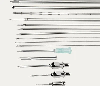
The diversification of diameter specifications reflects the refinement of clinical needs. The diameter of spinal needles is typically denoted by Gauge (G), with common specifications ranging from 22G to 29G. Larger-gauge needles (e.g., 22G) have a larger inner diameter and faster cerebrospinal fluid (CSF) flow rates, making them suitable for diagnostic lumbar punctures requiring rapid collection of large sample volumes. Finer needles (e.g., 25G–29G) significantly reduce the incidence of post-dural puncture headache (PDPH), decreasing the rate from approximately 30% with traditional 22G needles to less than 5%. However, the corresponding CSF flow rate is slower, prolonging the procedure time. This trade-off prompts clinicians to select the most appropriate gauge based on specific requirements.
Length selection is equally important. Spinal needle lengths typically range from 3.5 inches to 7 inches (approximately 9–18 cm). Standard adult lumbar punctures often use 3.5-inch needles, while obese patients or those with anatomical abnormalities may require 5-inch or even longer needles. Pediatric needles are shorter, typically 1.5–2.5 inches. Length selection not only affects the success rate of the puncture but also relates to operational safety; needles that are too long increase the risk of accidental injury, while those that are too short may lead to puncture failure.
Tip design is the core of a spinal needle's performance. Traditional cutting-point needles (Quincke needles) have a simple design, but they cut rather than separate dural fibers during puncture, resulting in larger dural defects, which is a major cause of PDPH. Modern pencil-point needles (e.g., Whitacre, Sprotte) feature a conical tip and a side aperture. They separate dural fibers rather than cut them, significantly reducing CSF leakage. This design reduces the incidence of PDPH to 1–2% and has become the preferred choice for many clinical scenarios.
Material innovation has also driven the development of spinal needles with special functions. Radiopaque needles incorporate barium or bismuth compounds into the shaft, allowing for clear visualization under fluoroscopy and improving the accuracy of intraspinal procedures. Thermosensitive needles integrate miniature temperature sensors, which can be used to monitor CSF temperature and assess spinal cord perfusion. These specialized needles play unique roles in complex clinical settings.
Bioabsorbable materials represent one of the future directions for spinal needle development. Experimental polylactic-co-glycolic acid (PLGA) needles can gradually degrade within the body, making them particularly suitable for situations requiring prolonged drainage and avoiding the need for secondary removal procedures. Although still in the experimental stage, this material demonstrates a new direction for the evolution of spinal needles.
Quality control permeates the entire process from material selection to the final product. Medical-grade stainless steel must comply with ASTM F138 standards to ensure biocompatibility. Each batch of material undergoes chemical composition analysis, mechanical property testing, and corrosion testing. Finished needles must pass sharpness tests, patency tests, breakage tests, and bioburden tests to ensure their safety and efficacy.
From a broader perspective, the material evolution of spinal needles reflects a universal rule in medical device development: progressing from meeting basic functional requirements to enhancing safety and patient comfort, and then developing specialized functions and personalized options. Each material innovation brings new possibilities to clinical practice, driving lumbar puncture from a high-risk procedure to a safe and routine diagnostic and therapeutic modality.