Procedure Of Ptc Percutaneous Hepatic Puncture Cholangiography
Nov 10, 2022
1. Transaxillary intercostal puncture
(1) The puncture approach is generally 8 ~ 9 ribs or 9 ~ 10 intercostal space in the right midaxillary line. When possible, it is better to observe the variation of the liver directly under X-ray monitoring, and adjust the height, direction and depth of the puncture point.
⑵ Disinfection, covering, local anesthesia at puncture site.
(3) Insert the needle according to the puncture point selected above, with the tip pointing to the tip of the xiphoid process in a horizontal direction.
In general, the injection is about 8 ~ 13cm, and the bile duct is thicker. When the needle is inserted into the bile duct, a breakthrough can be felt. At this point, the needle core is pulled out and replaced with a syringe. While slowly withdrawing the needle, suction is performed at the same time. If the bile is extracted, the withdrawal stops, indicating that the needle tip is in the bile duct. If the bile is not extracted, the puncture fails when the needle is withdrawn to 1/2 of the needle path. The needle should be withdrawn to the subcutaneous area and the direction should be changed slightly before puncture. Continue 4 ~ 5 times, still have not extracted bile should stop the operation, so as not to damage too much liver tissue.
⑸ can also use the next method, into the needle to the appropriate depth, first inject a small amount of contrast agent, in the X-ray screen display to judge the position of the needle. If the needle strays into the blood vessel, the contrast agent will be diluted and quickly flow away; If the needle is in the liver parenchyma, the contrast agent will remain motionless; If the contrast agent enters the hepatic biliary duct, the slow flow of the contrast agent to the hepatic hilum can be seen.
⑹ After the successful puncture, the needle was fixed, the syringe with plastic tube was connected, part of the bile was extracted and sent for bacterial culture; Then slowly inject 20ml of 30% ~ 50% of the warm meglumine. When the patient feels the liver area is slightly swollen, the injection should be stopped and the film should be taken. If the bile duct is highly dilated, the contrast dose can be appropriately increased.
After taking the tablet, try to suck out the bile mixed with contrast agent, so as not to leak the bile. If the photo is satisfactory, you can end the inspection. If not satisfied, the contrast agent can be injected again for imaging.
2. The puncture site of transabdominal puncture was selected under the right costal margin. The puncture point was 2cm below the xiphoid process and 2cm to the right of the midabdominal line. The puncture needle should be 12cm long. This method is suitable for patients with liver enlargement.
3. Transperitoneal puncture This method is performed through the bare area behind the liver. Because this naked area remains constant even when the liver is enlarged; And this puncture will not damage important organs, nor will biliary peritonitis or intraperitoneal bleeding occur. Right phrenic nerve block was performed before angiography. Methods: The anterior edge of sternocleidomastoid muscle 2 ~ 3cm above the right clavicle was raised with 2%, and the range of motion was reduced, indicating that phrenic nerve block was effective.
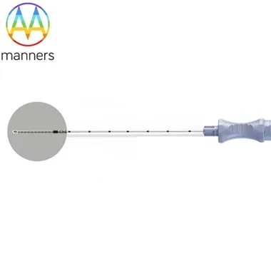
Then, the patient lay prone and underwent conventional local anesthesia at the upper margin of the right rib 11, 6 ~ 7cm away from the posterior median line. The liver was punctured with a puncture needle with a length of 15cm, with the needle slightly pointing upward and inward. When the needle was punctured 10 ~ 12cm, the needle was removed by the above method and bile was extracted to indicate successful puncture [Figure 1].
The procedure for injecting contrast agent and taking pictures is the same as before.
The success rate of this approach is much higher than that of transaxillary puncture.