A Standardized Operational System For Breast Fine Needle Aspiration
Jun 14, 2026
https://my.clevelandclinic.org/health/diagnostics/24204-breast-biopsy-overview
Keywords: Breast Fine Needle Aspiration, FNA, Quality Control, False Negative, Standardized Operation, Risk Management
Although Breast Fine Needle Aspiration (FNA) is a routine procedure, its diagnostic accuracy relies heavily on standardized execution and rigorous quality control. A failed FNA-resulting in unsatisfactory samples or false negatives-can lead to delayed diagnosis, unnecessary repeat biopsies, or even the loss of optimal treatment windows. Therefore, establishing a comprehensive quality management system spanning from pre-operative preparation to post-operative analysis is paramount.
Pre-Operative Phase: Credentialing and Equipment Assurance
- Personnel Qualification and Certification: Not every physician is qualified to perform FNA. Operators must undergo systematic training in anatomy, imaging, and puncture techniques. Ideally, the procedure should be performed by a breast specialist or a specially trained interventional radiologist. Regular simulation training and case assessments are necessary to ensure technical proficiency. Individual operator databases should be established to track satisfaction rates and diagnostic accuracy per procedure.
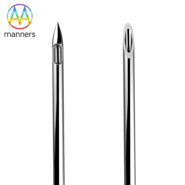
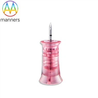
- Equipment and Consumables Management: Ensure that biopsy needles are within their expiration date and that packaging is intact. Needle tip sharpness is critical; reusable needles must undergo strict cleaning, lubrication, and sterilization protocols, with regular inspections for burrs or bends. Ultrasound equipment must be in optimal working order, with probe frequency, gain, and focal zones finely adjusted according to the depth and nature of the lesion.
Intra-Operative Phase: Technical Essentials and Real-Time Quality Control
- Precise Image Guidance: This is the cornerstone of successful FNA. The operator must confirm that the needle tip is within the lesion in two perpendicular planes. For cystic lesions, ensure the tip is centered within the cavity; for solid masses, move the needle tip within the mass to sample different areas. For microcalcifications, precise alignment with the calcification cluster is required.
- Immediate Sample Adequacy Assessment: This is the greatest advantage of FNA and its most critical quality control step. The operator or accompanying cytotechnician should immediately expel the aspirate onto glass slides for smearing, fixation, and rapid staining. A quick microscopic review should determine: ① Whether there is sufficient cellularity (typically requiring at least 6–8 groups of benign epithelial cells); ② Whether cell morphology is clear and free of crushing artifacts; ③ Whether representative components of the lesion are included (e.g., foam cells in cyst fluid, atypical cells in solid masses). If the sample is inadequate, immediate repeat aspiration is warranted.
- Specimen Handling and Labeling: After smearing, any remaining cellular material within the needle hub can be flushed into a cell preservative solution to create a cell block for immunohistochemistry and molecular testing. Every slide and tube must be labeled with a unique identifier containing the patient's name, medical record number, and puncture site to eliminate the risk of mix-ups.
Post-Operative Phase: Pathological Analysis and Result Feedback
- Standardized Pathological Reporting: FNA reports should adhere to internationally recognized classification systems (such as the IAC Yokohama System), clearly stating sample adequacy (satisfactory/unsatisfactory) and diagnostic categories (benign, atypical, suspicious for malignancy, malignant). For malignant cells, morphological features should be described whenever possible (e.g., ductal carcinoma, lobular carcinoma, mucinous carcinoma).
- Multidisciplinary Consultation and Follow-Up: Results that are inconclusive (e.g., atypical or suspicious) or discordant with imaging findings must be reviewed in a multidisciplinary team (MDT) meeting. For example, a highly suspicious BI-RADS 4C nodule with a "benign" FNA report is a "discordant" result. In such cases, blind faith in cytology should be avoided; instead, core needle biopsy or surgical excisional biopsy should be recommended for definitive diagnosis. Establish a robust follow-up system to monitor all FNA-negative patients with imaging for at least 12 months to rule out false negatives.
Risk Management and Complication Prevention
- Hemorrhage and Hematoma: The most common complication. Patients should discontinue anticoagulants prior to the procedure. Operators must avoid visible vessels during insertion. Apply firm compression for hemostasis post-procedure. Small hematomas can be observed for spontaneous resolution; large hematomas may require needle aspiration or drainage.
- Pneumothorax: Rare but serious. It is most common during biopsy of high axillary lymph nodes or deep lesions near the chest wall. Operators must be familiar with anatomical layers and keep the needle path parallel to the chest wall. If it occurs, emergency management is required.
- Infection and Needle Tract Seeding: Strict adherence to aseptic principles is mandatory. While there is a theoretical risk of tumor cell seeding along the needle tract, this risk is extremely low with FNA due to the fine gauge of the needle, and is significantly lower than that associated with core needle biopsy.
Conclusion
In summary, quality control in Breast Fine Needle Aspiration is a systematic project requiring close collaboration between clinicians, radiologists, and pathologists, integrating standardized operations into every step. Only by establishing a strict culture of quality control can the clinical value of FNA be maximized, ensuring patient safety and diagnostic reliability.