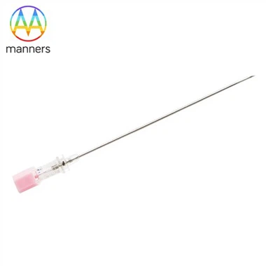
Ultrasound Echogenicity Enhancement Technology For EBUS-TBNA Needles
Jul 08, 2026
Why Laser-Etched Texturing is Crucial for Safe and Precise Ultrasound Guidance
https://profed.olympuschina.com/gs/thoracicsurgery/12628/
EBUS (convex probe, typically 7.5 MHz or 5-12 MHz variable frequency) enables real-time intraoperative visualization of airway walls, vessels, lymph nodes, and needle position. However, smooth metal surfaces reflect ultrasound almost entirely, rendering only a hairline hyperechoic artifact. This makes pinpointing the exact needle tip location and shaft trajectory difficult-particularly when puncturing deep lymph nodes or when the needle shaft aligns at shallow angles to the ultrasound beam ("needle tip loss"), elevating risks of vascular injury or over-penetration. Consequently, modern EBUS-TBNA needles require controlled surface micro-structuring to achieve reliable ultrasound echo enhancement (echogenicity enhancement).
Physical Principle: When ultrasound encounters a metal surface patterned with periodic micro-grooves or protrusions, it produces abundant scattered echoes alongside specular reflection. These scattered signals appear on the B-mode image as a broader, brighter hyperechoic band compared to a smooth surface. Operators can readily track the bright tip artifact and proximal shaft course. Microstructure depth, spacing, and arrangement dictate scattering intensity and directionality-insufficient depth yields weak enhancement, excessive depth creates stress concentrators or harbors contaminants.
Primary Implementation Methods:
- Laser Etching/Marking: Nanosecond/picosecond fiber lasers ablate helical grooves, annular dot matrices, or cross-hatched grids onto the needle shaft near the tip. Helical grooves provide consistent visibility regardless of rotational orientation, representing the preferred solution for premium EBUS needles (e.g., ViziShot2 series). Five-axis lasers achieve variable-pitch helices with ±0.01mm precision.
- Micro-Sandblasting + Chemical Etching Combinations: Sandblasting creates initial roughness, followed by localized laser marking for depth gauging and incidental echogenicity. Lower cost but compromised uniformity.
- Echogenic Coatings (Rare): Plasma-sprayed titanium oxide creates a roughened surface, but coating delamination risks limit application in disposable devices.
- Clinical Significance: Effective echogenic marking empowers operators to confirm: ① successful transmural puncture (disruption of the bright airway wall echo by the advancing needle tip artifact); ② intranodal needle tip position (bright tip artifact encased within the hypoechoic lymph node); ③ appropriate insertion depth and angle (estimating the length of the needle shaft within tissue). This proves especially critical for shortening the learning curve of novice operators and constitutes a mandatory imaging verification step during FDA/CE registration-typically requiring clear visualization of the needle tip and ≥5mm of the shaft across various incident angles (30°/45°/60°/90°) within tissue-mimicking phantoms.
During procurement, requesting manufacturer-provided ultrasound phantom imaging demonstrating the echogenicity pattern is advisable. Focus on the length of the marked zone (typically 10-20mm near the tip) and whether the instructions specify compatible ultrasound frequencies. Low-cost compatible needles lacking effective etching or relying solely on ink markings (which are sonolucent) pose significant safety hazards and are unsuitable for formal clinical use.