The Precision Mechanical Architecture Of The Veress Needle
Jun 18, 2026
https://en.wikipedia.org/wiki/Veress_needle
From Spring to Bevel: How the Six Components of a Veress Needle Work in Concert
Although the Veress needle appears to be a simple slender metal tube, it integrates several precision-engineered components-each playing an irreplaceable role in the high-risk maneuver of blind puncture and pneumoperitoneum establishment.
According to industry standards, the Veress needle typically measures 80–150 mm in total length, with an outer diameter (OD) of 2.5–5 mm and an inner lumen diameter of 1.5–3 mm. These dimensional parameters directly govern component design and mating tolerances.
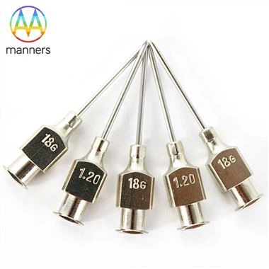
1. Outer Cannula (Cutting Cannula)
The outer cannula forms the main shaft of the Veress needle, usually made of medical-grade stainless steel (304 or 316L) with a tapered or streamlined profile to minimize insertion resistance.
Its distal end features a sharp bevel (typically 15°–30°) responsible for incising the abdominal wall layers.
The OD range (2.5–5 mm) dictates the trade-off between wall thickness and lumen size:
Excessively thin walls risk buckling
Excessively thick walls increase tissue trauma
High-quality cannulas are precision-ground to ensure bevel concentricity deviation < 0.01 mm; any eccentricity may cause directional drift during insertion.
2. Inner Stylet / Blunt Obturator (Blunt Tip Stylet)
This is the defining safety elementof the Veress needle.
The inner stylet is a blunt-ended rod, slightly smaller in diameter than the lumen, with a smoothly domed tip.
Resting state: The pre-compressed spring pushes the stylet 1–2 mm beyond the outer cannula tip, presenting a blunt profile.
During insertion: Abdominal wall resistance pushes the stylet back into the cannula, exposing the sharp bevel for cutting.
Upon peritoneal entry: Resistance vanishes → spring extends → stylet re-deploys, shielding the cutting edge and protecting intra-abdominal viscera.
This spring–stylet linkageis the fundamental distinction between a Veress needle and a conventional puncture needle.
3. Spring Mechanism (Spring Assembly)
The spring supplies the actuating force for stylet deployment. Usually housed within the hub and coaxial with the stylet shaft, it must be engineered to exacting specifications:
Too stiff: Stylet fails to retract → difficult penetration
Too weak: Delayed or incomplete deployment → loss of protective function
Industry standards require the spring to maintain stable force–displacement characteristics after thousands of compression cycles, across an operating temperature range of –20 °C to +50 °C.
Premium Veress needles undergo individual spring-force verification at final inspection.
4. Side Port(s) / Lateral Hole(s)
One or more lateral apertures are positioned 3–5 mm proximal to the distal tip. These-not the needle tip-are the primary gas outlet.
Rationale:
Prevents blockage when the tip contacts omentum or bowel
Avoids high-velocity focal CO₂ jets that could traumatize viscera
Side port diameter is typically 1.0–1.5 mm; some designs employ dual or helically arranged ports to optimize flow distribution and reduce localized pressure.
5. Hub & Stopcock Assembly
The hub is the surgeon's handling interface, commonly finished with anti-slip knurling, molded from medical polycarbonate or machined stainless steel. Internally it houses:
The spring preload/seating structure
A stopcock valve (2-way: ON/OFF; or 3-way: INSUFFLATE / OFF / VENT)
The hub terminates in a standard male Luer-Lock connector for insufflator tubing.
The assembly must be gas-tight, with a leak rate ≤ 0.1 mL/min at 15 mmHg.
6. Depth Markings & Color Coding
To aid insertion-depth estimation, the shaft is inscribed with etched or printed graduation rings (commonly every 1 cm). Many manufacturers apply color-coded hubs or shaft bands to denote length variants (e.g., green = 120 mm, blue = 150 mm). These markings also serve as visual reference points under fluoroscopy or CT when needed postoperatively.
The Value of Component Synergy
The six elements do not function in isolation. During a standard insertion:
Surgeon grips the hub and aligns the tip at the umbilical incision
Skin/fascia resistance compresses the spring → stylet retracts → bevel cuts
Peritoneal breach → resistance drops → spring deploys stylet
Side ports enter the peritoneal cavity
Stopcock opened → CO₂ flows through lumen and exits via side ports → pneumoperitoneum established
Failure of any single element-spring fatigue, side port occlusion, bevel dulling, stylet seizure-can result in failed entry or iatrogenic injury. Hence, finished Veress needles require full-functional testing of all subsystems, ensuring that every "click"heard at the bedside is both crisp and reliable.