The Art Of Millimeter Precision: Full Workflow Analysis And Quality Control System For Interstitial Needle Intervention
Apr 29, 2026
The Art of Millimeter Precision: Full Workflow Analysis and Quality Control System for Interstitial Needle Intervention
Precise, safe and effective interstitial needle application in cervical cancer treatment extends far beyond simple puncture and implantation. It constitutes a rigorous, interconnected systematic engineering integrating radiology, surgical techniques, radiation physics and nursing management - defined as the fine art of millimeter-scale precision. Negligence in any link triggers dose deviation, compromising therapeutic efficacy or increasing complication risks. This article comprehensively analyzes the full clinical workflow from pre-procedural planning to post-treatment evaluation, and establishes standardized quality control specifications.
I. Phase 1: Meticulous Preoperative Assessment and Virtual Planning
Sound preparation forms the cornerstone of reliable interstitial treatment. Most core design work is completed digitally before physical intervention.
1. Patient Selection and Informed Consent: Standardized indication screening (bulky tumor, eccentric growth, recurrence, etc.), general condition evaluation and anesthesia risk assessment. Medical teams fully explain procedural necessity, operational process, potential risks (bleeding, infection, perforation, pain) and clinical benefits, obtaining written informed consent.
2. High-Quality Imaging and Target Contouring: Pelvic high-resolution MRI scanning (combined with CT when necessary) in fixed treatment positioning. Radiation oncologists accurately delineate GTV, HR-CTV and all organs at risk including bladder, rectum, small intestine and sigmoid colon. Clear target boundary definition is the prerequisite for accurate needle deployment.
3. Virtual Pre-Planning: Physicists and clinicians conduct digital interstitial simulation on 3D treatment planning systems. Based on tumor morphology, they customize needle quantity, insertion trajectory, angle and depth, selecting matched auxiliary templates and access routes to achieve optimal preoperative dose coverage and organ protection.
II. Phase 2: Image-Guided Precise Implantation Surgery
This stage transforms virtual digital plans into clinical reality, routinely performed in operating rooms or dedicated brachytherapy suites under intravenous sedation or general anesthesia.
1. Position Fixation and Sterile Preparation: Strict lithotomy position consistent with imaging and treatment posture. Standard disinfection, draping and aseptic manipulation to reduce infectious risks.
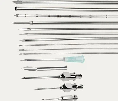
2. Applicator Placement: Intrauterine tandem insertion for combined protocols, followed by vaginal template fixation closely fitting the cervix and vaginal fornix. Grid-type template holes provide standardized parallel access channels to ensure uniform needle spacing.
3. Real-Time Image-Guided Puncture:
- Ultrasound Guidance: Transabdominal or transvaginal real-time monitoring tracks needle advancement, preventing excessive myometrial penetration or serosal perforation while avoiding visible large vessels.
- Combined Bimanual Examination: Tactile tissue resistance assessment verifies imaging findings for dual confirmation.
- Standardized Needle Insertion: Implant each needle strictly in accordance with virtual planning, recording insertion depth and template positioning data.
4. Post-Implant Fixation and Imaging Verification: Stable immobilization of needles and tandems via vaginal gauze and fixing devices to prevent intra-treatment displacement. Post-implant CT/MR scanning is mandatory to acquire accurate applicator and needle coordinates for final dose calculation.
III. Phase 3: Individualized Dose Optimization and Treatment Delivery Based on Actual Placement
1. Image Fusion and Structural Reconstruction: Coregister post-implant CT with preoperative high-definition MRI. Map clearly visualized needle coordinates from CT onto MRI with superior soft-tissue resolution, completing target and organ-at-risk re-contouring on fusion images.
2. Inverse Dose Optimization: The core technical advantage of modern brachytherapy. Import all needle dwell points into the planning system, setting clinical optimization goals (HR-CTV D90 > 85 Gy, rectal D2cc < 65 Gy, etc.). Algorithms automatically calculate individualized dwell time and source positioning to generate optimized conformal dose distribution. Oncologists and physicists jointly review dose-volume histograms and isodose curves for fine adjustment.
3. Treatment Implementation and Real-Time Monitoring: Transmit validated plans to the afterloading system. Transfer immobilized patients to the treatment suite and complete pipeline connection. The remote afterloading device delivers miniature radioactive sources sequentially to preset dwell positions for segmented irradiation. Whole-process video and voice monitoring ensure patient safety.
IV. Standardized Quality Control System: The Lifeline of Safety and Efficacy
1. Personnel Qualification and Training: Specialized certification and regular continuing education for radiation oncologists, medical physicists and radiographers, with standardized simulation training and skill assessment.
2. Equipment Quality Assurance: Periodic calibration of afterloading source positioning accuracy and timing precision; regular verification of CT/MRI geometric accuracy and image fusion reliability; sterile inspection and integrity testing of templates and interstitial needles.
3. Process Quality Management:
- Mandatory post-implant CT verification to ensure dose calculation based on authentic anatomical geometry.
- Independent secondary plan review by certified physicists to eliminate computational errors.
- Double-person verification of patient information, plan parameters and pipeline connection before treatment delivery.
4. Individualized Follow-Up and Data Management: Establish standardized medical records documenting needle quantity, positioning and dosimetric parameters. Long-term follow-up monitors local control, survival outcomes and late toxicities to support continuous technical optimization.
Conclusion
Conclusion
Interstitial brachytherapy for cervical cancer represents a multidisciplinary, millimeter-scale static stereotactic radiation surgery guided by precise imaging. As the core operational instrument, interstitial needles exert full clinical value only under standardized full-process quality control. Every procedure link - from virtual planning and precise implantation to image verification and inverse optimization - demands extreme accuracy. Beyond high-dose delivery, this rigorous system maximizes curative benefits and minimizes normal tissue damage. Mastering this millimeter-level artistic technique equips clinical teams with one of the most powerful and precise weapons in radiation oncology for managing complex cervical cancer cases.