Menghini Liver Biopsy: An In-Depth Analysis Of The Clinical Gold Standard And The Evolution Of Modern Technology
Apr 09, 2026
Liver Biopsy: An In-Depth Analysis of the Clinical "Gold Standard" and the Evolution of Modern Technology
The liver, the body's most complex metabolic and detoxification organ, presents a wide spectrum of diseases that are often asymptomatic in their early stages. Accurate diagnosis is the prerequisite for effective treatment of conditions ranging from viral hepatitis and fatty liver disease to cirrhosis and hepatocellular carcinoma. Among the numerous diagnostic tools, percutaneous liver biopsy has long been regarded as the "gold standard" for liver disease diagnosis, yet its name often evokes fear and misunderstanding in patients. This article systematically elaborates on the clinical value, technical principles, and risk assessment of liver biopsy. Combined with an analysis of the technological evolution of the modern Menghini liver biopsy needle, it explains how this critical diagnostic technique achieves a balance between high precision and minimal invasiveness, providing irreplaceable histological evidence for clinical decision-making.
I. The Clinical Positioning of Liver Biopsy: The "Microscopic Court" Transcending Serology and Imaging
While serological and imaging tests provide a "functional report" and a "morphological snapshot" of the liver, they have inherent limitations:
* Liver Function Tests (LFTs): Only reflect the biochemical results of hepatocyte damage or cholestasis, and cannot distinguish the specific pathological stages of inflammation, fibrosis, or steatosis.
* Imaging Examinations: Ultrasound, CT, and MRI can detect space-occupying lesions and assess tissue texture, but lack cellular-level resolution for diffuse diseases (e.g., grading inflammation in non-alcoholic steatohepatitis or staging early liver fibrosis).
The core value of liver biopsy lies in providing a "pathological verdict":
1. Grading Inflammatory Activity: Determines the degree of inflammation in chronic hepatitis, guiding the intensity of antiviral therapy.
2. Staging Fibrosis: Quantifies the progression of liver fibrosis from F0 (none) to F4 (cirrhosis), which is crucial for prognosis assessment and determining treatment windows.
3. Differentiating Steatosis from Steatohepatitis: Distinguishes simple fatty liver from steatohepatitis accompanied by inflammation and necrosis, the latter carrying a significantly higher risk of progression to cirrhosis.
4. Characterizing Space-Occupying Lesions: Serves as the definitive basis for differentiating hepatocellular carcinoma, cholangiocarcinoma, hemangioma, and focal nodular hyperplasia.
5. Diagnosing Metabolic and Hereditary Liver Diseases: The diagnosis of conditions like Wilson's disease and hereditary hemochromatosis relies on special staining and quantitative analysis of copper/iron in liver tissue.
II. The Technological Evolution of Liver Biopsy: From "Blind Puncture" to Intelligent Guidance
Liver biopsy has evolved from an early, experience-based "blind puncture" technique into a precise interventional procedure guided by multimodal imaging.
* Puncture Guidance Techniques:
* Real-time Ultrasound Guidance: Has become the standard configuration. High-frequency probes can clearly display the needle tract, allowing real-time avoidance of major intrahepatic blood vessels, bile ducts, and the gallbladder, minimizing the risks of bleeding and bile leakage.
* CT Guidance: Used for lesions in特殊 locations poorly visualized by ultrasound, or for biopsies requiring极高 spatial precision prior to procedures like radiofrequency ablation.
* Ultrasound Elastography Fusion Guidance: Prior to biopsy, shear wave elastography is used for a preliminary assessment of liver stiffness, allowing targeted selection of the most suspicious area for puncture, thereby improving the diagnostic yield of the sample.
* Innovations in Puncture Pathways:
* Classical Percutaneous Pathway: The most commonly used, suitable for most diffuse liver diseases and peripheral lesions.
* Transjugular Intrahepatic Approach: Suitable for patients with severe coagulation disorders, massive ascites, or a history of liver transplantation. The biopsy needle enters the hepatic vein via the jugular vein and inferior vena cava, then punctures from the hepatic vein wall into the liver parenchyma. Bleeding via this route flows into the venous system, significantly reducing the risk of intra-abdominal hemorrhage.
* Laparoscopic or Intraoperative Direct-Vision Biopsy: Used for特殊 complex cases or exploration of multiple lesions.
III. The Engineering of Biopsy Needles: The Design Philosophy of the Menghini Needle and Modern Customization
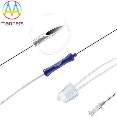
Since its introduction in 1958, the classic design of the Menghini liver biopsy needle-a cannula system with a retractable sample notch-remains one of the most widely used biopsy needle types globally. Its workflow perfectly embodies the balance between reliability and safety.
1. Classical Two-Step Workflow:
* Step One: Cannula Puncture. A cannula with a sharp stylet penetrates the skin, subcutaneous tissue, and liver capsule, reaching the target position.
* Step Two: Sample Notch Exposure and Tissue Cutting. The stylet is partially or completely withdrawn, exposing the elongated sample notch on the side of the cannula within the liver tissue. Subsequently, while maintaining negative pressure (usually via an attached syringe), the cannula is rapidly rotated and advanced, using its sharp cutting edge to sever and capture the liver tissue strip lodged in the notch.
2. Engineering Advantages of the Menghini Needle:
* Tissue Integrity: Can obtain continuous, intact liver tissue strips up to 1.5-2.5 cm in length, meeting the sample volume requirements for routine pathology, special stains, and molecular testing.
* Operational Standardization: The fixed procedure is easy to learn and master, ensuring a high success rate among different operators.
* Safety: The cannula design can temporarily compress the needle tract during puncture, and the sample notch can be re-sealed by the stylet after sampling, theoretically reducing the risks of needle tract seeding and bleeding.
3. Modern Customization Evolution:Advanced manufacturers like Manners Technology, building upon the classic Menghini design, offer deep customization solutions to meet individualized clinical needs:
* Needle Gauge Customization: Offers specifications from 16G to 20G. Finer needles (e.g., 18G) cause less trauma and pain, suitable for children or patients with borderline coagulation function; while 16G needles can obtain thicker tissue strips, providing richer diagnostic information.
* Needle Length and Stroke Customization: The effective working length and sample notch exposure stroke can be customized based on patient physique, liver size, and lesion depth, enabling precise depth control.
* Needle Tip Geometry Optimization: Modifies the bevel angle and sharpness of the stylet tip to reduce the "breaking sensation" and discomfort at the liver capsule; optimizes the micro-serration design of the cannula's cutting edge to ensure a single cut yields a complete sample, avoiding "crush artifact."
* Ergonomic Handle: Employs handles that conform to grip mechanics, are slip-resistant, and feature ultrasound-visible markers, enhancing operational stability and visibility under ultrasound.
IV. Safety and Perioperative Management: Systematic Risk Control
The safety of liver biopsy is founded upon strict patient selection, standardized operation, and systematic monitoring.
* Preoperative Assessment Checklist:
* Coagulation function and platelet count are mandatory tests; severe abnormalities are absolute contraindications.
* Imaging evaluation to明确 confirm the absence of high-risk lesions like hemangiomas or hydatid cysts in the planned puncture path.
* Patient education, including proper breathing training (puncture is typically performed during end-expiration breath-hold) to ensure cooperation.
* Intraoperative Safety Protocols:
* "One-Stick-One-Check" Principle: After each puncture, immediately observe the needle tract under ultrasound for any active bleeding.
* Control of Sample Number: 1-2 passes are usually sufficient for diffuse diseases, avoiding unnecessary multiple punctures.
* Postoperative Monitoring and Complication Management:
* Spectrum of Complications: The incidence of minor complications (local pain, transient hypotension) is approximately 5%; the incidence of serious complications (bleeding requiring transfusion or intervention, bile leak, pneumothorax, accidental puncture of adjacent organs) is below 0.5%.
* Standardized Monitoring: Absolute bed rest for 4-6 hours post-procedure with vital sign monitoring, followed by gradual resumption of activity. Bleeding is the primary risk, most often occurring within 2-6 hours after the procedure.
* Contraindications:
* Uncooperative patient or inability to breath-hold.
* Uncorrectable severe coagulation dysfunction.
* Clinical or imaging suspicion of liver hemangioma or hydatid disease.
* Uncontrolled massive ascites.
* Acute infection in the right pleural cavity, lung, or biliary system.
V. Future Outlook: The Reshaping Role of Liver Biopsy in the Era of Precision Medicine
Even as non-invasive diagnostic technologies (e.g., serum markers, radiomics, AI-assisted image analysis) advance rapidly, the "gold standard" status of liver biopsy remains irreplaceable in the short term. However, its role is evolving from a "screening tool" to a "platform for precise verification and molecular diagnosis."
1. The Cornerstone of Molecular Pathology: The obtained liver tissue is not only for H&E staining but is also precious material for immunohistochemistry, gene sequencing, and proteomic analysis. For example, detecting specific gene mutations (e.g., TERT promoter, TP53) in hepatocellular carcinoma can provide crucial guidance for targeted and immunotherapy.
2. Guiding the Calibration of Non-Invasive Techniques: The histological staging obtained from liver biopsy serves as the "anchoring" standard for developing and validating the accuracy of non-invasive technologies like transient elastography and serum fibrosis models.
3. Trend of Technological Integration: Future biopsy procedures may integrate optical coherence tomography, Raman spectroscopy, and other real-time in-situ pathology techniques, allowing preliminary analysis of tissue性质 at the moment of puncture, achieving "biopsy-diagnosis" integration.
Conclusion
Liver biopsy, a technology spanning over half a century, has evolved into a highly precise, proceduralized, and predictable routine interventional procedure through continuous innovations in imaging guidance, the refinement of biopsy needles (e.g., modern customized Menghini needles), and strict perioperative management. It is not the endpoint of treatment but the key that unlocks precise, individualized liver disease management. With a full understanding of its clinical value, technical principles, and safety assurances, physicians and patients can jointly make the most informed diagnostic and therapeutic decisions, allowing this "liver detective" to reveal the essential truth of the disease at the most critical moments