Avf Needle:Farewell To Steel Needles, Embracing Change: A Detailed Standardized Protocol For The Puncture And Comprehensive Management Of Arteriovenous Fistula Soft Catheter Needles For Hemodialysis
Apr 09, 2026
Farewell to Steel Needles, Embracing Change: A Detailed Standardized Protocol for the Puncture and Comprehensive Management of Arteriovenous Fistula Soft Catheter Needles for Hemodialysis
With the evolution of hemodialysis treatment concepts and vascular access technology, the Dedicated Indwelling Soft Catheter Needle for Arteriovenous Fistula is progressively replacing traditional metal puncture needles, becoming a core instrument for maintaining the "lifeline" of dialysis patients. The "Expert Consensus on Arteriovenous Fistula Soft Catheter Needle Puncture"issued by the Chinese Nursing Association in February 2026, with an authority coefficient as high as 0.99, provides a solid scientific basis for the standardized application of this technique. This article aims to provide a comprehensive and systematic interpretation of the clinical application, operational standards, safety management, and training system for soft catheter needles, to promote the standardization of hemodialysis puncture techniques.
I. Core Technological Innovation: The Engineering Advantages and Physiological Protection Mechanisms of Soft Catheter Needles
1. Revolutionary Breakthrough in Instrument Design
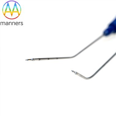
The design of the AV fistula soft catheter needle is a typical example of "clinical problem-oriented" engineering. Its core structure includes:
Inner Puncture Stylet: Made of high-strength, sharp-tipped medical-grade stainless steel or cobalt-chromium alloy, responsible for guiding the puncture.
Outer Indwelling Catheter: An ultra-thin, flexible soft tube made of polytetrafluoroethylene (PTFE) or biocompatible modified polyurethane. Its surface often has a hydrophilic coating to reduce friction, and the distal end features a multi-side-hole design (typically 3-4 holes) to ensure adequate blood flow and dialysis efficiency while reducing the risk of catheter wall apposition.
Integrated Connection System: Includes a transparent hub, a screw-on Luer connector, and a reflux-preventing heparin cap, forming a closed, needle-stick injury prevention system.
2. Biomechanical Protection Mechanism for Vessels
The rigid structure of traditional steel needles is the physical basis for repeated puncture injury. The mechanical advantages of the soft catheter needle are reflected in:
Reduced Shear Stress on the Vessel Wall: The flexible catheter can conform to the natural curvature of the vessel, reducing rigid friction. In vitro simulations and clinical observations indicate it can reduce mechanical micro-injury to the vascular intima by approximately 60%, which is especially critical for fragile vessels in patients with diabetes or the elderly.
Elimination of the "Piston Effect": Traditional steel needles create a "piston" motion within the vessel during patient movement, causing repeated vessel wall damage. Once indwelled, the soft catheter needle conforms to the vessel wall, eliminating this physical motion, thereby significantly lowering the incidence of vascular stenosis and pseudoaneurysm.
Optimized Hemodynamics: The multi-side-hole design disperses the impact of high-velocity blood flow, reducing shear-induced platelet activation and coagulation, theoretically lowering the risk of local thrombosis.
II. Refined Management Strategies for Clinical Indications and Contraindications
1. Stratified Indication Matrix
The application of soft catheter needles has moved beyond the realm of "can be used" to the stage of "best suited for" selection.
|
Applicable Population Category |
Specific Clinical Scenarios |
Core Benefits |
Level of Evidence & Consensus |
|---|---|---|---|
|
Strongly Recommended |
1. High-position fistulas, small vessel diameter, deep course. |
1. Flexibility reduces vascular puncture and indwelling injury. |
1A, 100% |
|
Extended Application |
1. Low-flow continuous renal replacement therapy with blood flow rates of 100-150 mL/min. |
1. Provides stable blood flow even at low rates, avoiding treatment interruption due to insufficient flow. |
1B, 100% |
2. Risk-Oriented, Graded Management of Contraindications
Absolute Contraindications: These are the "red lines" for technical safety that must not be crossed, including:
Active infection of the fistula vessel (local redness, swelling, heat, pain, or systemic signs of infection).
Complete thrombosis or occlusion of the fistula, preventing effective extracorporeal circulation.
Severe ischemia in the limb with the fistula (e.g., steal syndrome, resting pain).
Relative Contraindications: May be considered with caution only after thorough risk assessment and preparation of contingency plans:
Markedly thin vessel wall, large pseudoaneurysm (>2 cm diameter), or vascular malformation.
Breaks in skin integrity, dermatitis, psoriasis, eczema around the puncture site; skin issues must be addressed first.
Presence of severe coagulation disorders (e.g., INR >3.0, platelets <20×10⁹/L).
Extreme vessel tortuosity or severe calcification leaving no suitable puncture site on ultrasound assessment.
Key Decision Points: Relative contraindications are not absolute prohibitions but signals for decision-making escalation. The principle is "individualized assessment + pre-positioned contingency planning." For example, for a patient with a coagulation disorder, prepare local hemostatic agents beforehand and assign the most experienced nurse; for a patient with a large aneurysm, puncture can be performed under ultrasound guidance to avoid the aneurysm sac.
III. Evidence-Based Practice of the Standardized Operational Procedure
1. Pre-puncture Preparation and Assessment: From "Experience" to "Evidence"
Environment and Infection Control: Environmental disinfection is the first barrier against infection. The GB15982-2012 Class III environment standard is the baseline, with reinforcement of surface disinfection. Operators must strictly perform hand hygiene ("five moments") and wear sterile gloves.
Pain Management: For pain-sensitive patients, applying a lidocaine-prilocaine cream to the puncture site 30-60 minutes in advance (with a dedicated occlusive dressing) can effectively alleviate pain, improve patient experience, and enhance cooperation.
Ultrasound-Guided Vessel Assessment: For difficult-to-access vessels, ultrasound is the "gold standard." It is necessary to assess and record vessel inner diameter, depth from skin, blood flow direction, presence of stenosis/thrombus, and accurately mark the puncture site, vessel course, and depth reference line on the skin with a surgical marker.
2. Disinfection and Puncture: Evidence-Based, Precise Operation
Choice and Protocol of Disinfectant: The consensus clearly recommends evidence-based disinfection protocols, with the core principle being sufficient contact time. In clinical practice, 10% povidone-iodine is often the first choice due to its broad-spectrum and persistent effect, but adequate drying time must be ensured. When using chlorhexidine-alcohol, its rapid drying and persistent antimicrobial properties make it an efficient choice.
Use of Tourniquet: "When to Use" and "When Not to Use":
Use: In native arteriovenous fistula (AVF) and peripheral venous punctures, moderate use can distend the vessel, which is key to improving first-attempt success rate. Pressure should be moderate-enough to visibly distend the vessel without obliterating the arterial pulse-and the tourniquet must be released immediately upon successful puncture.
Do Not Use: In arteriovenous graft (AVG) and arterial punctures, tourniquet pressure can easily cause turbulence within the graft or a pressure gradient in the artery, increasing the risk of thrombosis and vessel injury; therefore, it is strictly contraindicated.
3. Refined Techniques for Puncture, Fixation, and Needle Removal
Angle of Insertion: An insertion angle of 30°–45° is the optimal balance of mechanics and anatomy. An overly shallow angle may cause the catheter to tunnel excessively in the subcutaneous tissue, injuring it; an overly steep angle increases the risk of transfixing the posterior vessel wall. During insertion, the tail of the metal hub must be stabilized to prevent it from retracting due to tissue resistance.
"Double Confirmation" of Successful Puncture: Blood return in the metal hub only indicates the stylet tip has entered the vessel lumen. It is essential to continue advancing until blood return is seen in the outer catheter (usually transparent) to confirm the soft catheter tip has entered the vessel. This is the core step to avoid the scenario of "only the stylet tip is in" leading to indwelling failure.
The "Golden Rules" for Catheter Advancement: During advancement, either the "fix stylet, advance catheter" or "retract stylet 1 cm, then advance catheter" method can be used, but it is strictly forbidden to advance the catheter after the metal stylet has been completely withdrawn. Without its support, the soft catheter is highly prone to kinking or twisting. At least 5 mm of the catheter must remain outside the skin, and the connection must be secure. This is a critical safety measure to prevent the catheter from completely slipping into the vessel, a serious adverse event.
Three-Level Fixation Method:
Primary Fixation: Use a transparent, breathable, waterproof transparent dressing, applied tension-free with the puncture site as the center, allowing for easy observation.
Secondary Fixation: Use flexible adhesive tape to secure the catheter hub and connection tubing using the hollow elevation method or U-shaped fixation, buffering tension.
Tertiary Fixation: Secure the extracorporeal bloodlines to the patient's limb using a spiral or U-shaped method, then reinforce the entire setup with an elastic bandage or specialized fixation sleeve. Throughout, it is crucial to avoid any tape or dressing creating a circumferential constriction on the vessel.
Standardized Needle Removal and Disposal:
"Point Pressure": After needle removal, apply pressure with a sterile gauze roll 0.5-1 cm proximal to the puncture site, perpendicularly, with enough force to occlude blood flow but not completely obliterate the arterial pulse. Compression time: 15-30 minutes for native AVF, 10-15 minutes for AVG. The punctured limb should not be used for blood pressure measurement or lifting heavy objects within 24 hours.
Sharps Safety Management: Must use soft catheter needles with an automatically activating safety shield. After the metal stylet is withdrawn, the safety shield should lock automatically or manually, and the stylet must be immediately and directly disposed of in a sharps container. Recapping with two hands is strictly prohibited in any form.
IV. Quality Assurance, Staff Training, and Safety Management System
1. Standardized Training and Qualification Certification
The adoption of soft catheter needles must be preceded by a standardized training system. A complete pathway including theoretical instruction, manikin simulation, clinical preceptorship, and independent assessment should be established. Upon passing the assessment, institutional operating credentials should be granted, with regular refresher training. The instructor team should consist of both vascular access physicians and hemodialysis specialist nurses.
2. Continuous Quality Monitoring and Adverse Event Reporting
Establish key performance indicators (KPIs) related to soft catheter needle puncture, such as first-attempt success rate, unplanned catheter removal rate, hematoma rate, infection rate. Any adverse event related to the soft catheter needle (e.g., catheter fracture, vessel injury, infection) must be entered into an adverse event reporting system for root cause analysis and process optimization.
3. Patient Education and Self-Management
Provide thorough education to patients and their families, covering activity guidance during catheter dwell, observation points (e.g., oozing, pain, swelling), shower protection methods, emergency management, etc. This is a crucial link in achieving a closed-loop safety management system both inside and outside the hospital.
Conclusion
The application of AV fistula soft catheter needles marks a significant step in hemodialysis vascular access management, transitioning from "rough puncture" to "precise protection." Its value lies not only in the replacement of a device but in driving a systematic upgrade of the entire nursing operation process towards evidence-based, standardized, refined, and patient-centered care. By strictly implementing the expert consensus and constructing a complete system encompassing training, operation, monitoring, and education, we can truly safeguard the "lifeline" of dialysis patients, ushering in a new era of high-quality, long-lasting vascular access management as we bid farewell to steel needles.