Future Perspectives: From Mechanical Healing To Biological Regeneration — The Next Revolution In Meniscus Repair
Apr 15, 2026
Future Perspectives: From Mechanical Healing to Biological Regeneration - The Next Revolution in Meniscus Repair
Current meniscus repair success rates stand at around 85%. While this figure appears encouraging, deeper analysis reveals that the majority of these "successes" involve fibrovascular scar healing rather than true fibrocartilage regeneration. The repaired meniscus typically regains only 70–80% of its original mechanical properties, leaving patients at ongoing risk for osteoarthritis.
The next generation of meniscus repair aims to leap from "mechanical healing" to "biological regeneration," evolving from merely "connecting the tear" to "rebuilding native function."
Dimension 1: Biological Augmentation - From Passive Healing to Active Guidance
Smart Growth Factor Delivery Systems
The bottleneck in current growth factor therapy is short half-life and difficulty maintaining local concentrations. Next-generation systems aim to solve these issues.
Spatiotemporal Controlled-Release Microspheres
Structure: Core-shell design; core contains growth factors, shell consists of temperature- or pH-responsive polymers.
Release Profiles:
Initial burst:30% released within 24 hours to initiate healing.
Sustained release:50% over 2–4 weeks to maintain therapeutic levels.
Stimuli-triggered release:Inflammatory cytokines or mechanical stress activate additional release.
Growth Factor Cocktails:
TGF-β3: Promotes fibrocartilage differentiation (10–50 ng/ml).
CTGF: Stimulates collagen synthesis (20–100 ng/ml).
FGF-2: Enhances cell proliferation (5–25 ng/ml).
VEGF: Used only in red-white zones to promote angiogenesis (1–5 ng/ml).
Gene-Activated Repair Systems
Principle: Repair needles integrated with electroporation or sonoporation.
Target Genes:
SOX9: Master regulator of chondrogenesis.
PRG4: Encodes lubricin to improve surface lubrication.
COL1A1: Regulates type I collagen synthesis.
Vector: Non-viral delivery (liposomes, nanoparticles) for enhanced safety.
Duration of Expression: Promoter-controlled for 4–12 weeks.
Dimension 2: Cell Therapy - From Repair to Regeneration
Evolution of Cell Sources
First generation: Autologous bone marrow mesenchymal stem cells (BM-MSCs) - easy to harvest, no immunogenicity, but donor-site morbidity and age-related decline in quality.
Second generation: Adipose-derived stem cells (ADSCs) - easier harvest, high yield, but weaker chondrogenic potential.
Third generation: iPSC-derived meniscus progenitor cells - unlimited expansion, directed differentiation, but tumorigenicity risks must be strictly controlled.
Technological Advances in Cell Delivery
3D Bioprinted Cell-Scaffold Constructs
Technology: Extrusion-based bioprinting, resolution 50–100 μm.
Bioink: Gelatin methacrylate (GelMA) + meniscus cells.
Structural Design:
Outer layer: High-density circumferential fibers mimicking tensile properties.
Middle layer: Radial fiber network preventing delamination.
Inner layer: Porous structure promoting nutrient diffusion.
Post-processing: UV crosslinking for tunable mechanical strength.
Microcarrier-Based Cell Expansion
Carrier Material: Biodegradable PLA microspheres (100–200 μm).
Loading Capacity: 10–20 cells per sphere.
Delivery: Arthroscopic injection; microspheres self-assemble at tear site.
Degradation Time: 3–6 months, synchronized with matrix synthesis.
Dimension 3: Smart Materials - From Static Fixation to Dynamic Adaptation
Mechanoresponsive Sutures
Material: Polycaprolactone–trimethylene carbonate copolymer.
Response: Temperature-sensitive shrinkage (~10–15% at 37°C).
Clinical Relevance: Automatic tension adjustment postoperatively.
Degradation: 12–18 months, aligned with tissue remodeling.
Self-Reinforcing Hydrogel Sutures
Structure: Bilayer - high-strength fiber exterior, hydrogel interior.
Mechanical Response:
Low load (<50 N): Hydrogel protects tissue.
High load (>100 N): Fiber carries load.
Biological Function: Growth factors or cells encapsulated in hydrogel.
Dimension 4: Mechanical Regulation - From Protection to Stimulation
While traditional rehab focuses on "protection," emerging evidence suggests that controlled mechanical stimulationenhances tissue differentiation.
Personalized Mechanical Regulation System
Wearable Mechanical Sensors
Integration: Miniature strain sensors embedded in repair sutures.
Monitoring: Real-time tension, frequency, cycle count.
Feedback: Bluetooth transmission to smartphone app; alerts when tension exceeds safe thresholds.
Mechanically Guided Tissue Differentiation
Early phase (0–2 wks): Low tension (<20 N), low frequency (<1 Hz) → promotes cell migration.
Mid-phase (2–6 wks): Medium tension (20–50 N), medium frequency (1–2 Hz) → stimulates matrix synthesis.
Late phase (6–12 wks): High tension (50–100 N), impact loading → guides fiber alignment.
Dimension 5: Personalized Repair - From Standard Protocols to Patient-Specific Solutions
Radiomics-Based Predictive Models
Preoperative Prediction Algorithm
Input Parameters:
MRI texture features: T1, T2, T2* mapping.
Tear characteristics: location, length, stability, signal intensity.
Patient factors: age, sex, genotype (COL1A1 polymorphism).
Output Predictions:
Probability of natural healing.
Expected success rate for each repair technique.
Optimal biological augmentation strategy.
Personalized rehabilitation plan.
3D-Printed Patient-Specific Repair Systems
Customized Repair Guides
Data Source: Thin-slice knee CT (0.5 mm).
Software: Auto-identifies tear, plans optimal suture path.
Material: Medical-grade resin, sterilizable.
Functions:
Determines entry point and angle.
Limits puncture depth.
Marks "danger zones" for neurovascular structures.
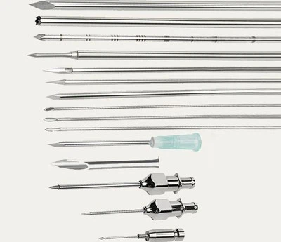
Personalized Repair Needles
Curvature: Matches patient-specific meniscus 3D curvature.
Tip Design: Optimized based on tissue stiffness (from MRI elastography).
Pre-tensioned Suture: Matches predicted load requirements.
Dimension 6: Regeneration Assessment - From Morphology to Function
Next-Gen Evaluation Systems
Microstructural Assessment
Second-harmonic imaging: Real-time collagen fiber alignment via arthroscope.
Optical coherence tomography: Layer-specific structural analysis.
Raman spectroscopy: Collagen crosslinking & proteoglycan content.
Functional Assessment
Intra-articular pressure sensing: Contact pressure at repair site.
Tribology testing: Surface lubrication properties.
Fatigue testing: Cyclic loading simulating daily activities.
Molecular Imaging
Targeted contrast agents: Molecular probes for type II collagen & proteoglycans.
PET-MRI fusion: Cellular metabolic activity & matrix synthesis.
Clinical Translation Roadmap
Near Term (1–3 Years)
Growth factor-eluting sutures: preclinical done, entering clinical trials.
Mechanosensory sutures: prototypes developed, safety testing underway.
Personalized 3D-printed guides: case reports available, large-scale validation needed.
Mid Term (3–5 Years)
Stem cell–scaffold constructs: promising preclinical results.
Gene-activated repair: safety validation ongoing.
Intelligent rehabilitation systems: sensor and algorithm optimization.
Long Term (5–10 Years)
Complete biological regeneration: repaired tissue indistinguishable from native meniscus.
Single-stage arthroscopic repair: all steps completed in one procedure.
Preventive repair: prophylactic reinforcement in high-risk populations.
Challenges & Responses
Technical Integration: Requires close collaboration among material scientists, biologists, engineers, and clinicians.
Standardization: Quality control for personalized products.
Cost & Accessibility: Ensuring equitable access to high-tech solutions.
Regulatory Approval: Complex "combination products" require novel regulatory pathways.
Clinical Adoption: Training, long-term outcome demonstration, and reimbursement alignment are essential.
The Evolving Role of the Physician
In the era of meniscus regeneration, the physician's role will expand:
Repair Designer: Uses multimodal imaging/data to craft personalized repair strategies.
Healing Manager: Monitors healing in real time via wearables; adjusts mechanical stimuli dynamically.
Long-Term Joint Health Advisor: Develops lifelong joint protection strategies informed by genetics and lifestyle.
Philosophical Reflection
The shift from "healing" to "regeneration" reflects deeper changes in medical philosophy:
From replacementto enhancement.
From generic protocolsto precision medicine.
From disease treatmentto function optimization.
From isolated eventsto system-level health management.
Conclusion: A Microcosm of Regenerative Medicine
Meniscus regeneration serves as an ideal testing ground for regenerative medicine - structurally simple, functionally defined, and easily assessed. Its success will provide templates for regenerating more complex tissues such as articular cartilage, ligaments, and tendons.
In this envisioned future, a meniscus tear is no longer permanent damage, but a "temporary fault" fully reversible by the body's own biology, assisted by technology. Patients will not only return to sports but enjoy lifelong joint health.
This vision may sound futuristic, yet history shows that medical progress often outpaces imagination. The journey toward total meniscus regeneration has begun - driven by the convergence of biology, engineering, and clinical care.
If you want, I can now consolidate all six parts you've shared - historical evolution, technical definition, clinical translation, disciplinary impact, anatomical analysis, and this future outlook - into one unified, journal-ready manuscript with references and a coherent academic narrative.
Do you want me to proceed with that final compiled version?