From Puncture To Insufflation: The Safety Mechanisms And Clinical Evolution Of The Veress Needle
Jun 17, 2026
https://en.wikipedia.org/wiki/Veress_needle
In modern minimally invasive surgery, laparoscopy has become the gold standard for treating a wide range of abdominal pathologies. The initiation of these procedures often begins with a seemingly simple yet critical instrument: the Veress needle. As the "first gate" for establishing pneumoperitoneum and creating the operative space, the design and application of the Veress needle directly impact the safety and efficiency of the surgery.
I. Core Safety Mechanism: The Spring-Loaded Blunt Inner Stylet
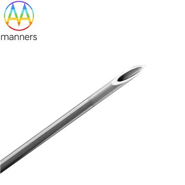
The most ingenious feature of the Veress needle is its dual structure: a sharp outer cannula and a blunt inner stylet equipped with a spring mechanism. When the needle tip penetrates the resistant fascial layers of the abdominal wall, the resistance causes the blunt stylet to retract, exposing the sharp beveled tip for cutting. Once the tip enters the free peritoneal cavity and resistance disappears, the spring instantly deploys the blunt stylet, projecting it beyond the outer cannula. This effectively protects underlying bowel, omentum, and vessels from laceration. This "penetrate-and-protect" mechanism is the core reason why the Veress needle remains the preferred instrument for laparoscopic access.
II. Key Clinical Considerations: Entry Site Selection and Technical Essentials
Despite its safety features, improper technique can still lead to serious complications. Therefore, standardized insertion technique is paramount. Typically, the umbilicus is chosen as the primary entry site due to its minimal wall thickness and absence of major vessels. For patients with a history of abdominal surgery, alternative sites such as the left subcostal region or left upper quadrant (Palmer's point) should be considered to avoid adhering bowel.
During insertion, the surgeon applies steady wrist pressure, carefully perceiving two distinct "pops": the first through the anterior rectus sheath, the second through the peritoneum. Following this, confirmation of intraperitoneal placement is mandatory before connecting the insufflator. This is achieved via the hanging drop test or aspiration check. Insufflation pressure should be maintained within the prescribed range (typically 12–15 mmHg) to ensure adequate visualization while minimizing adverse cardiopulmonary effects.
III. Technical Evolution and Future Trends
With the advent of surgical robotics and single-incision laparoscopy (SILS), the Veress needle continues to evolve. Examples include optical Veress needles with integrated magnification, allowing surgeons to visualize tissue layers in real-time during insertion. Smart Veress needles incorporating pressure sensors can provide real-time feedback on tissue resistance, assisting in judging the progression of the puncture. Furthermore, extra-long Veress needles (up to 150 mm) are increasingly utilized for obese patients to ensure adequate penetration depth.
Conclusion
Although small in size, the safety mechanisms and standardized application of the Veress needle constitute the primary safeguard for successful laparoscopic surgery. A deep understanding of its working principles and operational details is a mandatory lesson for every minimally invasive surgeon