From Intracavitary Application To Needle-Sculpted Dosimetry: How Interstitial Needles Reshape Dose Boundaries in Cervical Cancer Brachytherapy
Apr 29, 2026
From Intracavitary Application to Needle-Sculpted Dosimetry: How Interstitial Needles Reshape Dose Boundaries in Cervical Cancer Brachytherapy
In definitive radiotherapy for cervical cancer, brachytherapy (afterloading) holds an irreplaceable position. Its physical cornerstone - the inverse square law - endows it with the unique capability to precisely target tumors while sparing adjacent normal tissues. Traditional intracavitary afterloading, via intrauterine tandems and vaginal applicators, delivers favorable dose distribution to the cervix and surrounding tissues. However, for locally advanced disease, large tumors (>4 cm in diameter), irregular lesions (barrel-shaped, endophytic growth), or eccentric masses with parametrial invasion, the limitations of pure intracavitary approaches become prominent. It fails to fully cover peripheral tumor margins, creating cold spots that lead to local treatment failure.
Under such circumstances, Interstitial Brachytherapy (ISBT), centered on its core instrument - the interstitial needle - evolves from an adjuvant technique into a decisive factor for therapeutic success. It marks a paradigm shift in cervical cancer afterloading: from passive intracavitary treatment confined to natural anatomical cavities, to active dose sculpting guided by precise needle deployment.
I. Interstitial Needles: Dose Brushes Breaking Anatomical Restrictions
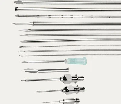
Interstitial needles are hollow, slender metal cannulas designed to serve as temporary channels for radioactive sources within and at the margins of tumor target volumes. Unlike intracavitary applicators restricted to the geometric centers of the uterine cavity and vagina, they can be accurately inserted into any high-dose required region based on three-dimensionally contoured tumor targets (GTV, HR-CTV).
1. Genuine 3D Conformity: Traditional intracavitary dose distribution presents a pear-shaped pattern centered on the uterine tandem. Interstitial needles enable physicians to sculpt highly conformal dose fields matching irregular tumor geometry by adjusting needle quantity, insertion angle and depth. For unilateral parametrial invasion, oblique needle placement directly pushes high-dose coverage to tumor edges.
2. Eliminating Dose Cold Spots: Distant tumor regions (anterior/posterior cervical lips) suffer severe dose attenuation with sole intracavitary irradiation. Implanting 1–2 interstitial needles within these marginal lesions establishes secondary radiation sources, elevating local dose and eradicating cold spots. This physical mechanism raises the local control rate of locally advanced cervical cancer from 60–70% to over 85%.
II. Technological Evolution of Interstitial Needles: From Blind Manual Puncture to Image Guidance
Clinical efficacy heavily relies on insertion accuracy, representing a continuous upgrade of imaging navigation systems.
1. 2D X-Ray Fluoroscopy Era: Early interstitial insertion depended on pelvic examination and fluoroscopic imaging. Operators implanted needles manually or via vaginal templates based on anatomical experience, with dose planning formulated on 2D radiographs. This method carried high uncertainty, unable to assess the 3D spatial relationship among needles, tumors, bladder and rectum.
2. CT/MR 3D Image-Guided Era (3D-IGBT): The current gold standard. Pre-procedural CT or superior MR imaging delineates clear boundaries of tumors, uterus, bladder, rectum and small intestine. Real-time intraoperative ultrasound guidance ensures accurate needle positioning and vascular avoidance. Post-implant CT/MR verification is mandatory to confirm actual needle placement. Subsequent 3D dose optimization visually balances tumor coverage and organ-at-risk tolerance, with interstitial needles acting as a physical bridge connecting pre-treatment planning and precise post-implant dose calculation.
III. Design and Selection of Interstitial Needles: A Diversified Armamentarium for Individual Scenarios
Modern interstitial needles form a systematic portfolio tailored to diverse clinical demands.
1. By Material & Structure
- Stainless Steel Cannula: The most widely used option. Graduated hollow shafts facilitate depth control; partial models with lateral side holes allow segmented source dwellage for complex modulated dose distribution.
- Plastic Catheter System: A flexible alternative. A sharp puncture trocar creates the access channel, followed by soft indwelling plastic catheter placement for improved patient comfort and stable fixation.
2. By Insertion Route & Auxiliary Tools
- Transvaginal Insertion: The primary approach. Standardized vaginal templates feature evenly spaced grid holes to ensure parallel, equidistant needle placement, enhancing geometric reproducibility for vaginal and parametrial lesions.
- Transperineal / Transabdominal Insertion: For complex cases. Indicated for extensive pelvic invasion, vaginal stenosis or recurrent tumors, requiring high-precision CT/ultrasound navigation and customized dose planning.
3. Combined Applicator Strategy: For most advanced cases, tandem combined with interstitial needles serves as the optimal solution. The intrauterine tandem covers central cervical and lower uterine doses, while peripheral needles extend high-dose coverage to invasive margins, achieving centralized radical irradiation plus peripheral marginal containment through synergistic plan optimization.
IV. Quantitative Clinical Benefits: From Local Control to Survival Improvement
High-level clinical evidence validates the value of interstitial needle technology:
- Large-scale international multicenter trials including the EMBRACE study confirm that MRI-guided 3D interstitial brachytherapy elevates the 3-year local control rate of locally advanced cervical cancer above 90%, while restricting grade ≥3 severe toxicities (rectal ulcer, vesicovaginal fistula) below 5%.
- For bulky initial tumors, interstitial technique is the only reliable method to deliver radical dose (HR-CTV D90 ≥ 85 Gy) while protecting critical organs.
- In salvage treatment for recurrent cervical cancer, especially central pelvic recurrence, interstitial brachytherapy avoids extensive pelvic exenteration and offers curative radiotherapy opportunities.
Conclusion
The seemingly simple interstitial metal needle stands as the ultimate dose-sculpting tool in modern precision cervical cancer radiotherapy. It liberates radiation oncologists from anatomical limitations of natural cavities, enabling active high-dose field modulation and customized coverage for irregular tumors. Advances from 2D empirical puncture to 3D intelligent image guidance directly translate to superior local control, prolonged survival and preserved quality of life. As an indispensable dose sculptor balancing radical tumor eradication and functional protection, interstitial needles define the upper boundary of comprehensive therapeutic outcomes in cervical cancer radiotherapy.