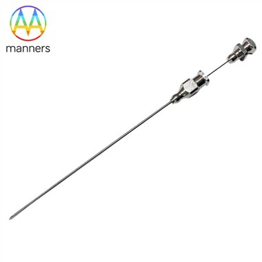
Bevel, Diamond Or Echo Tip: Choosing PTC Needle Geometry
Nov 11, 2026
The Pain Point: Two Needles, Same Gauge, Different Outcomes
Two 18G needles from different boxes can behave like different instruments. One enters a fibrotic duct wall cleanly and holds its line; the other slides off the wall, deflects along the capsule, and leaves the operator convinced that the duct moved. The variable is not gauge. It is tip geometry, together with the surface finish of the cannula and the stiffness of the shaft behind it.
This distinction matters because tip geometry is usually the last thing considered when a tray is opened. Selection frequently defaults to whatever configuration the kit contains, and the consequences - a deflected pass, a wall that recoils, a tip that cannot be seen on ultrasound - are attributed to anatomy or to operator technique rather than to the instrument. In a procedure where a single millimetre of deflection determines whether a drainage catheter can be placed, that attribution error is expensive.
How Tip Geometry Interacts with Tissue
Every needle tip concentrates force. The shape of that concentration defines what happens at the moment of contact with a duct wall. A beveled tip presents a single sharp leading edge and concentrates force along a line. It cuts rather than pushes, produces a clean linear track, and gives the operator a distinct, comparatively sudden loss of resistance on entry. The trade-off is directional bias: the bevel tends to deflect the tip away from the leading edge as it advances, so a beveled needle in soft tissue will curve unless the shaft is stiff enough to resist it.
A diamond or conical tip distributes force radially and symmetrically. It dilates rather than cuts, which is advantageous against a thin, mobile duct wall that would otherwise be pushed away from a cutting edge. It also produces less directional bias, so the path tracks the shaft more faithfully. The cost is higher penetration force and a less distinct entry signal - the operator feels gradual resistance change rather than a clear transition.
An echogenic tip is a manufacturing treatment rather than a distinct geometry: the tip surface is modified so that it reflects ultrasound strongly, usually by dimpling, coating, or a defined geometric feature. The benefit is continuous sonographic visibility of the tip, which matters most in the sub-3 mm duct, where the tip has to be watched into the target rather than felt into it. Echogenicity can be combined with either beveled or diamond geometry.
Surface finish completes the picture. Polishing, plating, electroplating, and ultrasonic cleaning affect friction along the track. A smoother, well-finished cannula reduces the insertion force required and reduces the tendency of the needle to drag surrounding tissue with it, which in turn reduces the small parenchymal injuries that later present as bleeding. Custom fabrication to a 2D or 3D drawing or a supplied sample allows tip angle, bevel length, and shaft stiffness to be specified together rather than independently.
Classifying Needle Tip Options for Biliary Access
Beveled tips in standard bevel, short bevel, and long bevel configurations cover most PTCD work. Long bevels cut a longer track and are useful for thickened, fibrotic duct walls in chronically obstructed systems. Short bevels provide more precise control and are preferred when the duct is close to a vessel that must not be crossed.
Diamond and trocar-type tips are used where the wall is thin and mobile, and in transjugular or transhepatic approaches where the target must be dilated rather than incised. They are also selected when the operator wants a straighter path in soft, fatty liver.
Echo-enhanced tips are chosen when ultrasound is the primary modality and the target is small, or when the operator needs to confirm tip position independently of fluoroscopy - a useful redundancy when contrast has already been injected.
Fine needles in the 21G–22G range are available in all three families and are the practical default for non-dilated ducts. Standard 18G–19G needles concentrate the selection decision on tip geometry, since gauge is fixed by the need for wire support. Large-bore introducer systems in the upper size range incorporate their own tip designs and are selected for drainage capacity rather than for puncture behaviour.
A Practical Guide to Tip Selection
Assess the wall before choosing the tip. A chronically obstructed duct with a thickened, rigid wall responds best to a cutting bevel. A thin-walled, minimally dilated duct responds best to a dilating diamond tip. This judgement can be made from the pre-procedure imaging in most cases and should be recorded in the plan.
Match tip to imaging modality. If the puncture will be performed under ultrasound in a small duct, echogenic tip visibility is not a luxury. If the puncture will be performed under fluoroscopy in a dilated duct, tip echogenicity is irrelevant and tip cut quality is not.
Fire the tip, do not push it. The commonest avoidable error is slow, continuous advancement, which allows the tip to deflect and the wall to recoil. Advance in short increments with an aspirating pause between them, so that entry is recognised at the moment it happens rather than after a centimetre of overshoot.
Aspirate after each increment. Bile return is the confirmation signal. Absence of return after a full anticipated depth indicates deflection; withdraw to the capsule and re-angle rather than advancing further on the assumption that the duct is deeper than expected.
Avoid dilating on an unconfirmed tip. If contrast injection shows a parenchymal blush rather than ductal filling, reposition. Dilation of a mispunctured track is the mechanism by which a small technical error becomes a bile leak that requires additional intervention.
Respect the bevel's bias. When using a beveled needle in soft liver, anticipate drift away from the leading edge and compensate at the capsule by adjusting the entry angle, or specify a stiffer shaft.
Field Experience: Selection Signals from Practice
In series comparing tip types for biliary access, the differences appear where the anatomy is difficult rather than where it is easy. Dilated ducts are forgiving and tend to succeed regardless of tip. Non-dilated ducts are where tip selection separates outcomes: echogenic, fine-gauge needles with a diamond or short bevel configuration consistently reduce the number of passes needed, because the tip can be tracked in real time rather than inferred from resistance.
A second recurring observation concerns fibrotic ducts in long-standing obstruction. Beveled tips shorten access time here, but only when the shaft is stiff enough to resist deflection; a flexible shaft behind a cutting bevel produces a curved path that misses the target even though the tip geometry is correct. The lesson is that tip and shaft must be specified together.
A third concerns finish. Cases complicated by minor oozing along the track are more frequent when insertion requires high force, which correlates with rougher surface finish and with needles that have been re-sheathed or handled roughly. Single-use handling and attention to surface specification are unglamorous but measurable.
Summary: Geometry Is a Clinical Decision
Tip geometry is not a manufacturing detail that happens to be visible on a datasheet. It determines whether the duct wall is cut, dilated, or pushed aside, and therefore whether the first pass succeeds. A bevel cuts and biases; a diamond dilates and tracks; an echogenic treatment makes the tip visible where visibility is the limiting factor. Surface finish governs friction, and shaft stiffness governs whether the tip's intended behaviour is realised. Selecting on gauge alone leaves the most consequential decision unmade.
Outlook: Specifying Tips Together with Shafts
The direction of travel for PTC instrument design is toward combined specification rather than isolated features: tip geometry matched to documented wall characteristics, surface finish reported as a friction-relevant parameter rather than a cosmetic one, and echogenic treatment offered as standard on fine-gauge needles rather than as an option. For procurement, the practical implication is to request needle samples for bench testing against representative tissue models before standardising a tray, and to keep at least two tip configurations available so that the decision can be made on imaging findings rather than on inventory. As the evidence base for tip selection matures, the expectation should be that this choice is documented in the procedure note alongside gauge and approach - because a decision that is not recorded cannot be improved.
< img alt="news-1-1" src="/uploads/24240/news/202604241539186d0ff.png " />