Needle-Level Infection Control In PTCD
Nov 11, 2026
The Pain Point: The Infection That Was Already There
Bile duct infection after percutaneous transhepatic cholangiographic drainage is rarely a simple failure of antibiotic choice. In patients with malignant biliary obstruction, infection is often present before the patient reaches the interventional suite: the obstructed system is a stagnant reservoir, bacterial colonisation is common, and the patient's general resistance is depressed by jaundice, malnutrition, and malignancy itself. The procedure then adds a second opportunity - instruments crossing the skin, the hepatic parenchyma, and the bile duct wall in a single continuous pass.
Because the infection may be pre-existing, a post-procedural fever is easily attributed to the patient's underlying disease and treated expectantly. That attribution is the trap. Cholangitis after PTCD can progress to bacteraemia and sepsis within hours, and the window for effective intervention is short. Strict aseptic technique during the procedure is not a formality that can be relaxed when access is difficult; it is the only infection control measure entirely within the operator's control.
How Contamination Actually Enters the Biliary System
There are three routes. The first is direct inoculation: skin flora carried on the needle tip through the capsule and into the duct lumen. This is the route that aseptic technique addresses, and it is directly proportional to the number of needle passes - each pass is a fresh inoculation event. The second is intraluminal seeding from contrast injection into a colonised, obstructed segment under pressure. Injecting contrast forcefully into a system with a distal obstruction can force infected bile into the bloodstream through cholangiovenous reflux. The third is ascending infection along the drainage catheter in the days and weeks after the procedure, which is a maintenance issue rather than an access issue but is strongly influenced by whether the initial drainage was complete and unobstructed.
Access technique influences all three. Multiple passes raise the first. High-pressure injection raises the second. Incomplete initial drainage - leaving a system that remains obstructed despite a catheter in place - raises the third.
Equipment Factors That Affect Infection Risk
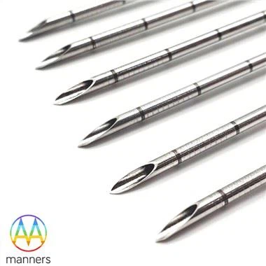
Needle configuration affects contamination in ways that are easy to overlook. Surface finish matters: a well-polished, plated, or ultrasonically cleaned cannula reduces friction and tissue drag, which reduces the trauma and micro-haematoma that provide a substrate for bacterial proliferation along the track. A rougher surface, or a needle that requires repeated re-advancement, produces more track disruption.
Tip geometry matters indirectly. A tip that requires multiple attempts to enter the duct multiplies inoculation events. A tip that enters cleanly on the first pass converts a multi-event contamination risk into a single one.
Cannula design and hub connections matter for the injection phase. Needles that allow controlled, low-pressure contrast injection - through a correctly sized lumen with a secure luer connection - reduce the risk of cholangiovenous reflux compared with forceful injection through a narrow lumen with a loose connection.
Kit composition matters for exchange count. Every additional instrument exchange in a contaminated field is an opportunity for the sterile field to break down. Trays that supply matched needle, wire, dilator, and catheter components reduce the number of times the operator reaches outside the prepared field.
Fine needles in the 21G–22G range reduce parenchymal trauma and therefore reduce the tissue substrate available for infection, but they do not reduce the need for asepsis. Large-bore introducer systems increase trauma per pass; their use should be justified by drainage requirements rather than by convenience.
A Practical Guide to Aseptic Access
Prepare the field as if the case will be difficult, because the difficult case is when asepsis fails. Full sterile draping, cap, mask, sterile gown, and gloves; skin preparation with an appropriate antiseptic allowed to dry fully; and a clear separation between the prepared field and the ultrasound probe, which should be covered with a sterile sheath and sterile gel.
Limit passes by planning them. Every pass should have a defined purpose. If a pass does not produce bile or contrast confirmation, stop, withdraw to the capsule, and re-plan on imaging rather than re-advancing in a new direction from depth.
Inject gently and confirm drainage. Use small-volume contrast injection under fluoroscopic observation. If the system fills and does not drain, the priority is establishing drainage, not obtaining a complete cholangiogram. A complete, pressurised cholangiogram in an infected obstructed system is a recognised trigger for cholangitis.
Use prophylactic antibiotics in the obstructed, previously infected system, and continue according to local protocol. Antibiotic prophylaxis does not substitute for technique; it reduces the consequence of the contamination that technique fails to prevent.
Drain adequately at the first attempt. Incomplete drainage leaves a residual obstructed segment and is a leading contributor to delayed infection. Verify catheter position and side-hole distribution before finishing the case.
Follow the post-procedural routine. Fasting for two hours, then a low-fat, high-protein, high-carbohydrate diet rich in vitamins, supports the patient's resistance during the period of highest risk. Monitor temperature and inflammatory markers in the first 48 hours rather than assuming a fever is disease-related.
Control instrument exchanges, because exchanges are where sterility fails. Each time a needle, wire, or dilator leaves the field and another enters, the connection between the patient's bile duct and the room is reopened. Practical measures are unglamorous and effective: prepare all components before puncture, position the tray so that the operator does not have to reach across the field, keep a second operator responsible solely for handling unused inventory, and avoid re-sheathing or re-using any component. Where a two-operator team is available, the assistant should open and hand off while the operator maintains the field. Where it is not, the sequence should be arranged so that no exchange requires the operator to turn away from the sterile area. Trays that arrive pre-arranged in the order of use reduce both exchange time and the temptation to improvise, and matched component sizing removes the most common reason for an unplanned extra instrument.
Field Experience: What Changes Outcomes
Centres that report low post-PTCD cholangitis rates tend to share three practices rather than three antibiotics. They limit needle passes by planning trajectory on pre-procedure imaging. They avoid full-pressure cholangiography before drainage is established. And they treat every post-procedural fever in the first 48 hours as procedure-related until proven otherwise.
A recurring error in difficult access is the pursuit of a perfect cholangiogram in a septic patient. The operator, having achieved access, injects to define anatomy and demonstrates a satisfying ductal map - and the patient becomes febrile within six hours. The anatomy could have been defined after drainage, at lower pressure, with less risk.
A second recurring pattern is access through a subcapsular or capsular haematoma created by an earlier pass. The haematoma is an excellent culture medium, and infection at that site can track along the catheter. This is one more reason to stop after a failed pass and re-plan rather than to persist in the same region.
Summary: Asepsis Is the Only Controllable Variable
Bile duct infection in malignant obstruction is frequently present before the procedure and cannot be eliminated by technique alone. What technique can do is avoid adding to it: minimise passes, keep the field sterile throughout every exchange, inject at low pressure, establish drainage before pursuing diagnostic imaging, and verify that drainage is complete. Needle surface finish, tip quality, and kit composition support these aims by reducing trauma and exchange count, but they cannot replace them. The operator who treats asepsis as a continuous requirement rather than an opening ritual controls the one variable that is fully within their power.
Outlook: Designing Trays for Infection Control
The next practical advance in PTCD infection control is likely to be tray design rather than pharmacology: single-pass access systems that reduce instrument exchanges, pre-assembled drainage sets that keep the sterile field intact, and specification of surface finish and lubricity as documented parameters. Suppliers who publish surface treatment and cleaning validation data alongside product specifications make it possible for procurement to select on infection-relevant criteria rather than on diameter alone. Combined with a standard of documenting pass count as a quality metric, this shifts infection control from individual vigilance to system design - the only level at which it can be reliably sustained.
< img alt="news-1-1" src="/uploads/24240/news/202604241539186d0ff.png " />