Ultrasound Visualization Adaptability Of Radioactive Particle Localization Needles
Oct 01, 2026
1. Industry Imaging Adaptation Pain Points
Traditional radioactive particle localization instruments have serious imaging adaptation defects in ultrasound-guided brachytherapy. Ordinary puncture needles have flat needle tip structure and weak ultrasonic reflection signal, resulting in blurred needle tip imaging, unclear puncture trajectory and difficult real-time positioning under ultrasound monitoring. Unoptimized needle body structure is prone to generate a large number of imaging artifacts and echo interference, covering tiny lesion targets and particle implantation points, leading to positioning deviation and inaccurate dose distribution. Traditional needles lack directional identification structure, and clinicians cannot judge the bevel tip orientation through imaging, easily causing unilateral particle offset during seed deployment. In addition, uneven surface processing and poor coating adhesion of traditional instruments cause unstable ultrasonic signal feedback, resulting in inconsistent imaging effects in different tissue layers, greatly reducing the accuracy and stability of ultrasound-guided particle implantation, and restricting the development of precise ultrasound intervention radiotherapy.
2. Core Ultrasound Visualization Working Principle
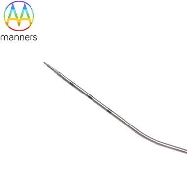
Radioactive Particle Localization Needles adopt exclusive echo-enhanced needle tip optimization design, which fundamentally solves the imaging blur problem of traditional localization needles. The specially treated needle tip structure strengthens ultrasonic wave reflection and echo signal intensity, realizing high-definition real-time display of needle tip position and puncture trajectory under conventional ultrasound equipment. The uniform and smooth needle body structure reduces scattered echo and artifact interference, ensuring clear imaging of target lesions and implantation areas without signal occlusion. Equipped with professional foil hub directional markers, it corresponds to the bevel tip angle one by one, enabling clinicians to accurately judge the needle tip direction through hub identification without relying solely on imaging. The high-precision laser scale calibration matches ultrasound depth measurement data, realizing accurate closed-loop control of puncture depth. Combined with low-resistance silicon coating and electropolishing smooth structure, it avoids tissue bleeding and exudation interference imaging, ensuring stable and reliable intraoperative ultrasound visualization effect.
3. Imaging Adaptive Product Classification
According to ultrasound visualization performance and directional positioning function, the products are divided into basic ultrasound visualization type and high-precision directional imaging type. The basic ultrasound visualization type adopts conventional echo-enhanced needle tip processing, with clear needle tip imaging and weak artifact interference, suitable for routine superficial tumor ultrasound-guided particle implantation and simple lesion localization. The high-precision directional imaging type optimizes needle tip echo structure and adds foil hub orientation marking system, with higher imaging resolution and accurate directional recognition function, specially suitable for deep tumor, multi-point particle implantation and complex anatomical position lesion precise localization. All products support customized optimization of echo intensity and structural parameters according to clinical imaging equipment differences, with complete size specifications and flexible customized configuration services.
4. Ultrasound-Guided Adaptive Operation Guidelines
Preoperative imaging calibration: Select matched visualization-grade localization needles according to ultrasound equipment resolution and lesion depth. Calibrate the echo effect of the needle tip in advance to confirm clear imaging and stable signal. Mark the tumor target range and particle implantation grid points under ultrasound multi-angle scanning, and formulate accurate puncture depth and angle scheme. Intraoperative imaging operation: Adjust ultrasound probe parameters to optimize imaging clarity, track the echo-enhanced needle tip trajectory in real time during puncture, and fine-tune the needle body angle according to lesion position. Judge the bevel tip orientation through the foil hub marker to ensure consistent seed deployment direction. After reaching the preset implantation point, stabilize the needle body to avoid imaging offset caused by shaking, and complete accurate seed release. Postoperative imaging verification: Scan the implantation area comprehensively through ultrasound, check the distribution position of radioactive particles, confirm no offset or missing implantation, and record imaging data for treatment effect evaluation.
5. Practical Imaging Adaptation Application Experience
Clinical ultrasound-guided brachytherapy practice fully verifies the excellent visualization adaptability of Radioactive Particle Localization Needles. Compared with traditional ordinary needles, the echo-enhanced products have 80% higher needle tip imaging clarity, real-time and intuitive puncture trajectory, effectively avoiding positioning errors caused by blurred imaging. The optimized structural design greatly reduces intraoperative artifact interference, ensuring that tiny lesions and multi-point implantation positions are fully displayed. The directional foil hub markers solve the long-standing industry problem of difficult judgment of needle tip direction, and the accuracy of particle directional deployment is significantly improved. The stable imaging adaptability can adapt to different tissue density environments such as glandular, muscular and adipose tissues, maintaining consistent high-definition imaging effect. The one-time accurate implantation success rate of ultrasound-guided surgery is increased from 85% to 99%, effectively improving the uniformity of radiotherapy dose distribution and clinical treatment effect.
6. Summary and Sublimation
Radioactive Particle Localization Needles effectively solve the industry pain points of weak ultrasonic signal, serious artifact interference and poor directional positioning ability of traditional tumor localization instruments. Through echo-enhanced structural optimization, directional marking design and precision processing upgrading, it realizes perfect adaptive matching with ultrasound-guided interventional surgery. It greatly improves the visualization degree, precision and stability of intraoperative particle implantation, avoids radiotherapy dose deviation and treatment failure caused by imaging errors, provides reliable technical support for ultrasound-guided precise tumor brachytherapy, and promotes the standardized and high-precision development of interventional radiotherapy imaging technology.
7. Industry Imaging Adaptation Development Suggestions
The industry should continue to iterate echo-enhanced technology for particle localization needles, develop high-sensitivity needle tip structures suitable for low-resolution ultrasound equipment, and expand the scope of equipment adaptation. Enterprises should strengthen the R&D of anti-artifact interference structural design to further improve intraoperative imaging purity. Medical institutions should formulate standardized ultrasound-guided particle implantation operation specifications based on product imaging characteristics, summarize and avoid common imaging positioning errors. Strengthen multi-center clinical imaging effect research, optimize product directional marking and depth calibration system, and comprehensively improve the industry's ultrasound precise localization technical level.