The Multi-Dimensional Application Value Of Breast Biopsy Needles In The Entire Cycle Management Of Breast Diseases
May 24, 2026
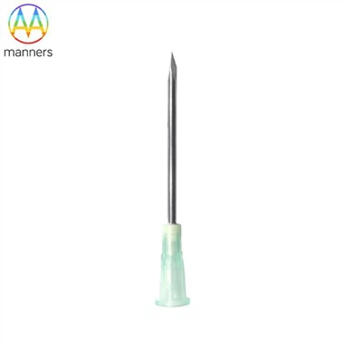
The breast biopsy needle, as a crucial interventional tool in the modern breast disease diagnosis and treatment system, has long transcended the simple scope of "taking a piece of tissue." It is an "intelligent appendage" that extends the perception and judgment capabilities of doctors. In the complete chain from screening discovery, precise diagnosis, treatment guidance to efficacy monitoring and scientific research exploration, it plays an indispensable multi-dimensional role. Manufacturers continuously iterate the design of the needle and the supporting technologies to enable it to handle various targets ranging from macroscopic masses to microscopic calcifications, from solid lesions to fluid cysts, greatly enriching the diagnostic arsenal of clinical doctors and profoundly changing the diagnosis and treatment path of breast diseases.
The Cornerstone of Precise Diagnosis: Addressing Diverse Imaging Findings
Mammography, ultrasound, and magnetic resonance imaging (MRI) are the three fundamental methods for detecting breast abnormalities. And the biopsy needle is the only way to convert these imaging "suspicions" into pathological "evidence."
For palpable or ultrasonically visible solid masses, ultrasound-guided core needle biopsy (CNB) is the standard method. The biopsy needle, under real-time ultrasound guidance, can precisely penetrate the interior of the mass and obtain one or more complete tissue strips. The samples obtained maintain the tissue structure, which is sufficient for diagnosing benign lesions (such as fibroadenoma), in situ carcinoma, or invasive cancer, and for performing histological grading. For clustered microcalcifications only visible on mammography, stereotactic biopsy is required. The patient lies supine on a specially designed biopsy table, the breast is compressed and fixed, and the mammography is used to perform three-dimensional positioning from two angles to guide the biopsy needle to accurately reach the calcium cluster area for sampling. The extracted tissue specimens need to be immediately subjected to mammography radiography to confirm that the calcification points have been successfully obtained, which is crucial for diagnosing early lesions such as ductal carcinoma in situ (DCIS).
When clinical examination reveals nipple discharge, especially single-channel bloody discharge, biopsy or brush sampling under the guidance of a ductoscope becomes possible. Finer specialized biopsy needles or brushes can directly sample the suspicious intraductal lesions through the working channel of the ductoscope, providing diagnostic evidence for intraductal papillomas or cancers. For inflammatory breast lesions or atypical diffuse changes, biopsy can help rule out inflammatory breast cancer or other specific inflammations and provide a clear diagnosis.
The Navigator of Treatment Decisions: From Pathology to Molecular Typing
In modern breast cancer treatment, the endpoint of diagnosis is no longer simply "benign" or "malignant." The tissue samples obtained through biopsy are valuable materials for a series of subsequent analyses and directly guide the formulation of treatment plans.
Through immunohistochemical (IHC) staining, the expression levels of estrogen receptor (ER), progesterone receptor (PR), HER2 protein, and Ki-67 proliferation index can be detected in biopsy samples, thereby classifying breast cancer into different molecular subtypes such as Luminal A type, Luminal B type, HER2 overexpression type, and triple-negative type. This classification serves as the fundamental basis for selecting endocrine therapy, anti-HER2 targeted therapy, or chemotherapy. Moreover, for patients who need to assess genomic risk (such as the 21-gene test by Oncotype DX) or genetic susceptibility (such as BRCA gene testing), sufficient biopsy tissue is also a prerequisite for conducting these expensive but crucial molecular tests. Manufacturers have directly supported this "one biopsy, comprehensive diagnosis" modern pathological model by providing core biopsy needles that can obtain higher-quality and more complete tissue sections, avoiding the need for patients to undergo a second invasive procedure due to insufficient samples.
A Window for Efficacy Evaluation and Scientific Exploration
The application of biopsy needles is not limited to the initial diagnosis. During the neoadjuvant treatment (chemotherapy, targeted therapy, etc. conducted before surgery), a second puncture biopsy of the primary lesion is performed at the mid-stage of the treatment to assess the tumor's response to the treatment (whether there is pathological complete response pCR or not), providing real-time evidence for whether to adjust the treatment plan. For patients with advanced breast cancer, when there is suspected drug resistance or new metastatic lesions, a biopsy of the metastatic lesion (re-biopsy) can be conducted to understand whether the molecular characteristics of the tumor have changed, thereby guiding the subsequent selection of precise treatment.
In the field of research, fresh tumor tissues obtained through biopsy are valuable resources for cutting-edge studies such as organoid cultivation, drug sensitivity testing, and single-cell sequencing. These studies contribute to a deeper understanding of tumor heterogeneity, resistance mechanisms, and the development of new treatment strategies. The biopsy technique that can efficiently and minimally invasively obtain high-quality live tissue samples serves as an important bridge connecting clinical and basic research.
Therefore, the products of breast biopsy needle manufacturers essentially provide clinicians with a powerful "information acquisition system." This system can convert the spatial positioning information from imaging into tissue specimens with complete biological information. From the initial qualitative diagnosis to the detailed molecular classification, and to the dynamic monitoring during the treatment process, the biopsy needle runs through the entire process. The manufacturer continuously optimizes the cutting efficiency of the needle, the integrity of sample preservation, and the ease of operation, thereby enhancing the efficacy and reliability of this "information converter," enabling clinical decisions to be based on increasingly solid and rich pathological evidence, and ultimately driving the diagnosis and treatment of breast diseases towards a more precise and individualized direction.