The Journey From A Single Needle To A Diagnostic Report
Jun 29, 2026
https://www.medicalexpo.com/medical-manufacturer/puncture-needle-3117.html
Core Angle: Revealing the entire process of how lung puncture samples are taken from the body to issuing a pathological report, focusing on the distinct values and technical challenges of cytological versus histological diagnosis.
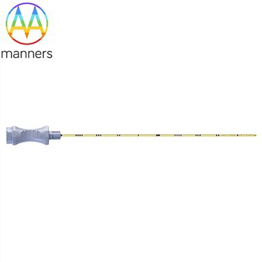
A slender puncture needle enters the lung, bringing out not just a few milliliters of tissue fluid or a small strip of tissue. This tiny specimen carries the "pathological code" that determines the patient's fate. From the needle tip to the microscope, and then to the final diagnostic report, it is a precise, rigorous, and challenging scientific journey.
I. Two Major Types of Puncture Specimens
- Fine Needle Aspiration Cytology (FNAC): Uses 22G-25G fine needles to obtain single cells or small clusters of cells via negative pressure aspiration. Advantages include minimal trauma, simple operation, and quick preliminary results (e.g., bedside rapid staining to determine presence of cancer cells). Disadvantages are that only cell morphology can be observed, tissue structure cannot be seen, differentiation capability for certain tumor subtypes (like adenocarcinoma vs. squamous cell carcinoma) is limited, and false negatives are prone to occur.
- Core Needle Biopsy (CNB): Uses 18G-20G cutting needles to obtain a complete tissue core (about 1-2 cm long, approx. 1 mm in diameter). This is currently the gold standard. It preserves intact tissue structure; pathologists can not only determine if cells are malignant but also assess their differentiation degree, depth of invasion, presence of vascular invasion, and perform immunohistochemistry and genetic testing, providing key evidence for targeted therapy and immunotherapy.
II. The "Decoding" Process in the Laboratory
- Fixation and Embedding: Specimens are immediately placed in formalin upon extraction to prevent cell autolysis. Subsequently, they are dehydrated, paraffin-embedded, and cut into thin sections of 4-5 micrometers.
- Staining: The most commonly used is H&E staining, rendering nuclei blue and cytoplasm red, displaying clear morphological features.
- Immunohistochemistry (IHC): This is the core technology of modern pathological diagnosis. Utilizing the principle of specific antigen-antibody binding, labeled antibodies are used to "seek out" specific proteins on the surface of tumor cells. For example, TTF-1 positivity indicates lung adenocarcinoma, p40 positivity indicates lung squamous cell carcinoma, and PD-L1 expression levels directly guide immunotherapy drug selection.
- Molecular Testing: DNA/RNA is extracted from tissue sections to detect driver gene mutations such as EGFR, ALK, ROS1, as well as immunotherapy-related indicators like MSI and TMB. This has ushered lung cancer treatment into an era of "individualized precision."
III. Challenges and Limitations Faced
- Insufficient Specimen Volume: When lung lesions are small or consist mostly of necrotic tissue, the obtained specimen may be insufficient to complete all necessary tests.
- Sampling Error: The puncture may only capture the edge or necrotic area of the tumor, failing to represent the characteristics of the entire lesion.
- Subjectivity of Manual Interpretation: Even experienced pathologists may have diagnostic disagreements when facing atypical cell morphologies. In recent years, AI-assisted pathological diagnosis systems are rapidly developing to help improve consistency and efficiency in interpretation.
Conclusion: Behind every lung puncture pathology report lies the concerted effort of radiologists, thoracic surgeons, and pathologists. This small puncture needle is not only a diagnostic tool but also a bridge connecting patients to precision treatment. It tells us: on the road against cancer, only by clearly seeing the true face of the enemy can we formulate the most effective battle plan.