The Evolution Of Technology: How Vacuum-Assisted Biopsy Needles Redefine The Precision Boundaries Of Breast Minimally Invasive Surgery
Apr 14, 2026
The Evolution of Technology: How Vacuum-Assisted Biopsy Needles Redefine the Precision Boundaries of Breast Minimally Invasive Surgery
Q&A Approach
When a breast lesion requires complete excision rather than mere sampling, how can the limitations of the traditional 14G core needle be overcome? How can the complete resection of benign lesions smaller than 3 cm be achieved while maintaining a tiny 3–5 mm incision? The advent of the Vacuum-Assisted Biopsy (VAB) system represents not merely an equipment upgrade, but a revolutionary leap in the philosophy of minimally invasive breast surgery. However, how does this system perform "millimeter-level sculpting" of lesions under real-time ultrasound monitoring?
Historical Evolution
Vacuum-assisted biopsy technology originated from advancements in breast imaging in the 1990s. In 1994, the U.S. FDA first approved directional vacuum-assisted biopsy devices for stereotactic biopsy of calcifications. In 1999, the development of ultrasound adapters expanded its application to ultrasound-guided procedures. The emergence of large-caliber 8G rotary cutting needles in 2005 made the complete resection of benign tumors feasible. After 2010, various rotary cutting needles (8G, 10G, 11G, 12G) formed a complete product matrix, catering to diverse needs from biopsy to excision. In 2017, the Breast Surgery Group of the Chinese Medical Association's Surgical Branch released an expert consensus, marking the entry of this technology into a stage of standardized application in China.
Technical Standard Definitions
The vacuum-assisted biopsy system is an integrated engineering solution:
|
System Component |
Technical Specification |
Functional Definition |
|---|---|---|
|
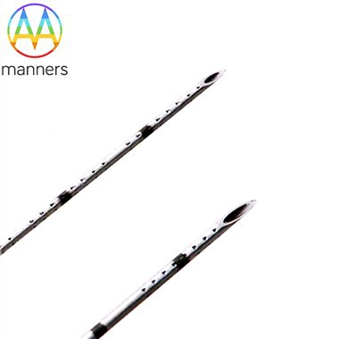
Rotary Cutter |
8–12G, Inner Diameter 2.3–3.2mm, Notch Length 20–25mm |
Core component for lesion capture and cutting |
|
Vacuum Pump |
Negative pressure 400–600 mmHg, adjustable |
Provides continuous suction to immobilize and aspirate tissue |
|
Drive System |
Rotation speed 300–1200 rpm, programmable |
Drives the rotation of the cutting blade |
|
Transport System |
Built-in delivery tubing, check valve design |
Transports cut tissue fragments out of the body intact |
|
Control Unit |
Foot pedal/Hand control dual mode, real-time pressure monitoring |
Allows operator precision control |
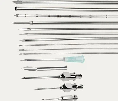
Essentials of Rotary Cutter Design
Needle selection strategies for different lesions:
12G Needle (2.7mm): Ideal for biopsy diagnosis; acquires contiguous samples; diagnostic accuracy >95% for lesions <1 cm.
10G Needle (3.0mm): Balanced model; suitable for excising 1–2 cm lesions, balancing efficiency and trauma.
8G Needle (3.2mm): Large caliber; suitable for complete resection of 2–3 cm benign tumors, though with a slightly higher risk of hematoma.
Operational Specifications Explained
The operational implications of consensus guidelines:
Anesthesia Strategy: Injection into the retromammary space is superior to perilesional infiltration, creating an operative space and reducing bleeding.
Needle Insertion Path: The needle track should be parallel to the long axis of the ultrasound probe to achieve full visualization.
Cutting Sequence: Begin fan-shaped cutting from the base of the lesion, moving upward progressively to ensure complete excision.
Hemostasis Technique: Residual cavity aspiration + surface compression for 15 minutes + pressure dressing for 24 hours.
Quality Control System
Key control points ensuring surgical safety:
Preoperative Verification: Precise three-axis measurement of the lesion via ultrasound to select the appropriate gauge.
Process Monitoring: Real-time ultrasound display of the cutter notch position to ensure it is entirely beneath the lesion.
Integrity Confirmation: Post-excision multi-plane ultrasound scanning to confirm no residual tissue.
Specimen Assessment: Intraoperative observation of specimen morphology to determine if it contains the entire lesion.
Complication Prevention and Control
Evidence-based risk management:
Bleeding Control: Dilute epinephrine solution (1:100,000–1:200,000) injected locally reduces hematoma incidence to <2%.
Skin Protection: Subcutaneous injection of tumescent fluid for superficial lesions establishes a safety buffer zone.
Chest Wall Avoidance: Maintain constant ultrasound monitoring of needle depth, keeping distance from the pectoralis major fascia >5 mm.
Infection Prevention: Strict aseptic technique keeps infection rates <0.1%.
Chinese Practice Data
Based on domestic multicenter studies:
Complete Excision Rate: 96.7% for benign tumors ≤2 cm; 89.5% for those ≤3 cm.
Diagnostic Accuracy: Sensitivity of 99.1% and specificity of 100% for malignant lesions.
Complication Rate: Hematoma 3.2%, skin injury 0.8%, pneumothorax 0.01%.
Cosmetic Satisfaction: Patient satisfaction score of 4.5/5.0 at 6 months post-op.
Expanding Technical Boundaries
New application scenarios for vacuum-assisted biopsy:
Neoadjuvant Response Evaluation: Multiple biopsies of the same lesion pre- and post-treatment to assess pathological response.
Management of Suspicious Calcifications: Complete excision of coarse calcifications visible on ultrasound.
Multifocal Lesions: Addressing up to 3 adjacent lesions through a single incision.
Male Breast Disease: Minimally invasive treatment of gynecomastia.
Future Technology Integration
Evolutionary directions for next-gen vacuum-assisted biopsy:
Real-time Pathological Feedback: Integrating Optical Coherence Tomography (OCT) at the needle tip to assess margins intraoperatively.
AI Navigation: Automatic identification of lesion boundaries and planning of optimal cutting paths.
Robotic Assistance: Robotic arms providing stable control, eliminating hand tremors.
Energy Platform Integration: Immediate radiofrequency hemostasis post-cutting to reduce hematomas.
As Professor Liu Yinhua, lead author of the consensus, stated: "Vacuum-assisted biopsy is not simply a larger bore needle, but a complete minimally invasive surgical system." From sampling to excision, from diagnosis to treatment, this technology is redrawing the precision boundaries of minimally invasive breast surgery.