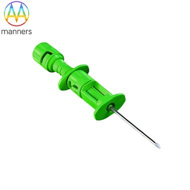
Techniques Of Bone Marrow Puncture And Biopsy in Non-Human Primates: Focus On Biopsy Needle Application
Aug 27, 2026
Non-human primates (NHPs) are core experimental animals for preclinical hematology research, stem cell drug development, immune mechanism exploration and translational medicine studies. High-quality, low-contamination and complete bone marrow specimens are the premise and foundation of reliable experimental data. Bone marrow puncture and core biopsy are the most mainstream in vivo bone marrow sampling techniques for non-human primates. Different from ordinary venous blood collection, this technology relies on professional hard hollow biopsy needles to penetrate the cortical bone of long bones and flat bones, and obtain qualified bone marrow fluid and intact bone marrow core tissues from the medullary cavity. The whole operation is highly dependent on the performance specifications, model matching and standardized application of bone marrow biopsy needles. Combined with mature animal experiment protocols and literature experience, this article systematically elaborates the full-process operation specifications of NHP bone marrow puncture and biopsy, focusing on the selection characteristics, application advantages and standardized operation essentials of dedicated biopsy needles, to provide standardized technical guidance for experimental researchers.
The basic technical principle of bone marrow puncture and biopsy in non-human primates is consistent with the classical canine bone marrow sampling system (Crow et al., 2009). The core logic is to use rigid hollow biopsy needles to break through the hard cortical bone barrier, accurately locate the medullary cavity, and obtain bone marrow fluid and solid core specimens through negative pressure aspiration and rotary cutting embedding. According to the anatomical characteristics of NHPs, the common sampling sites are divided into forelimb and hindlimb areas. The hindlimb priority sampling sites include iliac crest and femoral trochanteric fossa (Wolf & White, 2012), which are suitable for most medium and large-sized primate models; the forelimb alternative site is the humeral greater tubercle, with shallow anatomical position and convenient operation, suitable for supplementary sampling and repeated sampling experiments. Different sampling sites have differences in bone thickness, medullary cavity diameter and soft tissue coverage, which need to be matched with corresponding specifications of bone marrow biopsy needles to ensure sampling efficiency and specimen quality.
As an invasive bone operation, NHP bone marrow puncture and biopsy will cause definite tissue stimulation and pain response. To ensure animal welfare and stable experimental operation, all sampling operations must be performed under standardized anesthesia. Researchers can select general anesthesia or local targeted anesthesia according to the experimental cycle, sampling frequency and animal individual state, so as to achieve sufficient intraoperative and postoperative analgesia. In addition, the whole process must strictly abide by sterile experimental specifications, including preoperative shaving and skin preparation of the sampling area, multi-standard disinfection with povidone-iodine and alcohol, sterile towel laying, and wearing sterile gloves and surgical instruments, to effectively avoid local infection, wound inflammation and other postoperative complications that affect subsequent repeated experiments.
Classification, Selection and Performance Advantages of Special Bone Marrow Biopsy Needles
Bone marrow biopsy needle is the core precision instrument of NHP bone marrow sampling, which determines the success rate of one-time puncture, specimen integrity and peripheral blood contamination degree. At present, there are multiple mature commercial models of special bone marrow sampling needles for experimental animals, including Rosenthal, Klima, Illinois sternal/iliac needle and Jamshidi needle. Each model has differentiated design in needle length, gauge diameter, inner core structure and handheld grip, adapting to different animal body types, bone anatomical characteristics and experimental sampling purposes.
Among all needle types, the 14G Jamshidi bone marrow biopsy needle is the gold standard for NHP bone marrow core biopsy, which is the most recommended specification for experimental operations. This type of biopsy needle adopts optimized rigid hollow structure, with matched central solid stylet. The needle body has moderate hardness and toughness, which can effectively penetrate the cortical bone of primates without bending or slipping. The spiral cutting edge at the needle tip can stably embed and intercept complete bone marrow core tissues, avoiding specimen fragmentation and tissue extrusion damage caused by unprofessional needle types. Compared with thin-gauge aspiration needles, 14G Jamshidi biopsy needle has a larger inner cavity diameter, which can obtain sufficient-volume core specimens while ensuring safety, meeting the morphological detection, tissue sectioning and experimental index analysis needs of core biopsy. Compared with oversized thick needles, it can effectively reduce excessive bone trauma and intraoperative bleeding, balancing specimen quality and animal repeatable utilization.
All special bone marrow biopsy needles are designed with a matching central stylet, which is a key structural feature different from ordinary injection needles. The stylet can completely block the hollow cavity of the needle body during puncture, prevent skin soft tissue and bone debris from entering the needle cavity to block the pipeline, and ensure the smooth progress of subsequent negative pressure aspiration and core tissue cutting. In repeated sampling experiments, researchers need to select needle specifications according to animal body weight and bone size: short and fine-gauge biopsy needles for small primates (body weight less than 5kg), and conventional 14G medium-length Jamshidi needles for medium and large individuals, realizing precise matching of equipment and experimental objects.
Key Preoperative Precautions for Biopsy Needle Sampling
To avoid specimen coagulation, hemolysis and contamination, and improve the qualified rate of experimental samples, standardized pretreatment and operation specifications for biopsy needles must be implemented before sampling. First, heparin anticoagulation pretreatment of needles and syringes is mandatory. Before use, extract a small amount of heparin solution to flush the inner wall of the biopsy needle and the matched syringe, and completely discharge the residual liquid, forming a uniform heparin anticoagulant coating on the inner wall of the needle cavity. This operation can effectively prevent the rapid coagulation of bone marrow fluid after sampling, avoid the failure of smear preparation and cell detection caused by specimen coagulation, and is the key pretreatment step for high-quality bone marrow fluid acquisition.
Second, for long-term repeated sampling animal experiments, rotational selection of puncture sites should be adopted to avoid repeated puncture at a single site, which may lead to cortical bone hyperplasia, local tissue adhesion and increased puncture resistance, resulting in sampling failure and increased animal trauma. In terms of analgesia scheme, local analgesic injection into the skin and deep soft tissue of the puncture site is superior to simple general anesthesia, which can effectively relieve postoperative wound soreness and discomfort, reduce animal stress response, and facilitate postoperative recovery and subsequent repeated experiments.
In terms of sampling volume control, limited by the needle cavity capacity of the biopsy needle and the physiological blood volume of experimental animals, the sampling volume of a single puncture site shall not exceed 2mL. Excessive extraction will lead to serious peripheral blood dilution, reduce the proportion of nucleated cells and active bone marrow tissues in the specimen, interfere with experimental detection results, and affect the accuracy of hematological index analysis and tissue morphological observation.
Standard Operating Procedure for Iliac Crest Puncture (Core Biopsy Needle Operation)
Iliac crest is the most conventional and preferred sampling site for non-human primate bone marrow biopsy, with thick cortical bone, rich medullary cavity hematopoietic tissue and few adjacent important organs, featuring high safety and high sampling success rate. After the animal is fully anesthetized, place it in a prone position, fold both lower limbs under the abdomen to lift the hip bone and fully expose the iliac crest sampling area. Most non-human primates have sparse hair in the iliac crest area, which only needs simple shaving and skin preparation. The skin is disinfected alternately with povidone-iodine and alcohol, and a sterile hole towel is laid to isolate the surgical area and ensure sterile operation.
After confirming the iliac crest boundary by palpation, make a tiny skin incision with a sterile surgical blade at the anterior side of the iliac crest to reduce the friction resistance of the biopsy needle during penetration. Align the 14G Jamshidi biopsy needle with the incision, advance slowly to the anterior dorsal edge of the iliac crest, keep the needle body perpendicular to the iliac crest plane all the time, and apply continuous and stable downward pressure while rotating the needle body clockwise. The vertical needle insertion posture is the key to successful biopsy, which can ensure that the needle tip accurately enters the medullary cavity, avoid oblique penetration leading to cortical bone splitting and adjacent tissue damage, and maximize the cutting and sampling performance of the biopsy needle tip.
When the needle body penetrates the cortical bone, the operator will obviously feel the sudden reduction of penetration resistance, and the biopsy needle can be stably fixed in the bone tissue without shaking, indicating that the needle tip has successfully entered the medullary cavity. At this time, pull out the central stylet completely, connect a 10mL sterile syringe to the tail of the biopsy needle, and apply stable negative pressure by pulling the piston. Fresh primate bone marrow is dark red and foamy, with higher viscosity than peripheral blood, and contains tiny light-colored trabecular bone tissue, which is a qualified bone marrow specimen.
If no bone marrow fluid is extracted, do not pull out the needle body blindly. Rotate the biopsy needle 90°–180° to adjust the cutting and sampling range of the needle tip; if still invalid, reinsert the stylet, slightly adjust the depth of the needle body forward or backward, and apply negative pressure again. For diagnostic experimental specimens, the sampling volume of a single site is controlled within 1mL to ensure specimen purity; if a large volume of bone marrow samples is required for experiments, multiple puncture sites shall be selected for sampling instead of excessive extraction at a single site. After sampling, pull out the biopsy needle quickly, apply direct compression hemostasis on the puncture site for a long time, and suture the skin with single strand suture if persistent bleeding occurs.
Puncture and Biopsy Operation of Femoral Trochanteric Fossa and Humeral Proximal End
The femoral trochanteric fossa is an ideal supplementary sampling site for small non-human primates (body weight <5kg). The local bone structure is shallow, and the medullary cavity is concentrated, which is convenient for the positioning and penetration of fine-gauge biopsy needles. However, the proximal thigh muscle layer of large primates is thick, resulting in large puncture resistance and difficult operation. The core operation principle is consistent with iliac crest biopsy: place the animal in lateral position, hold the proximal thigh with the palm, locate the trochanteric fossa at the inner and distal side of the femoral greater trochanter by thumb palpation, insert the biopsy needle toward the femoral shaft to penetrate into the medullary cavity, and complete negative pressure aspiration and specimen collection according to the standard needle operation procedure.
The proximal humerus is another efficient alternative sampling site. The diameter of the humeral long bone is larger, and the medullary cavity is wider and shallower, making the biopsy needle positioning more accurate and the operation simpler. Place the animal in lateral position, hold the proximal forelimb, palpate the intertubercular sulcus between the humeral greater and lesser tubercles, insert the biopsy needle toward the humeral shaft to enter the medullary cavity. This site has low requirements for needle body precision, low puncture failure rate, and is suitable for rapid sampling and batch animal experiments.
Long Bone Perfusion Method and Core Biopsy Needle In-depth Application
Different from conventional single-needle puncture, the long bone perfusion method (Kushida et al., 2000) is a high-purity bone marrow sampling technology suitable for high-precision experiments, which requires the cooperative use of two bone marrow biopsy needles. The operation needs to insert matched biopsy needles into the medullary cavity at both ends of the long bone shaft respectively, inject sterile normal saline slowly through one biopsy needle, and collect the bone marrow-containing perfusate into a sterile closed container through the extension tube connected to the other needle. This dual-needle cooperative sampling mode effectively avoids peripheral blood contamination in conventional puncture, and the obtained bone marrow specimens have higher purity, which is suitable for stem cell separation, in vitro culture and high-precision omics detection experiments.
Differentiated Operation of Bone Marrow Aspiration and Core Biopsy Based on Biopsy Needles
Bone marrow aspiration and core biopsy share the same sampling site and biopsy needle equipment, but the postoperative needle operation logic is completely different, which determines the two types of experimental specimens: liquid bone marrow fluid and solid bone marrow core tissue. In bone marrow aspiration, after the biopsy needle is fixed in the medullary cavity, pull out the stylet and connect a syringe to apply negative pressure to extract liquid bone marrow fluid, which is used for cell counting, smear morphology and flow cytometry detection.
In bone marrow core biopsy focusing on tissue specimens, after pulling out the stylet, the Jamshidi biopsy needle is further advanced 1–2cm into the medullary cavity through stable pressure and slow rotation. The spiral cutting edge at the needle tip can completely wrap the bone marrow core tissue. Then loosen the needle body by rotating and swinging, slightly retract and reinsert to separate the bottom of the core tissue from the bone trabecula. After confirming that the tissue is completely separated, pull out the biopsy needle integrally through rotary traction. Finally, push the intact solid bone marrow core specimen out of the needle cavity with a linear pusher, and fix it with 10% formalin for tissue section and pathological analysis.
Conclusion
The standardized application of bone marrow biopsy needles is the core key to the success of non-human primate bone marrow puncture and biopsy experiments. The 14G Jamshidi biopsy needle, with its optimized structural design and stable sampling performance, is the preferred equipment for NHP core biopsy. Researchers need to select needle specifications according to animal body type and experimental purposes, strictly implement heparin pretreatment, sterile operation and site rotation protocols, and master the differentiated operation techniques of aspiration and core biopsy. Standardized needle selection and operation can effectively improve specimen purity and integrity, reduce animal trauma and experimental error, and provide solid technical support for preclinical hematology and translational medicine research based on non-human primate models.