Structure, Dimensions, And Sampling Efficiency Of Breast Core Biopsy Needle Tips
Jun 13, 2026
https://www.mayoclinic.org/tests-procedures/breast-biopsy/about/pac-20384812
The core of a breast biopsy needle is not merely a simple hollow tube, but a precisely engineered miniature surgical instrument. Its length, diameter (gauge), tip geometry, and sample channel design collectively determine whether sufficient, intact, and undistorted tissue samples can be obtained-essential for accurate pathological diagnosis.
Length and Diameter: The Golden Rule of Matching Lesion Depth
The length of the needle core is typically selected based on the depth of the lesion relative to the body surface, with common sizes including 10 cm, 15 cm, and 20 cm. A needle that is too short may fail to reach deep-seated lesions, while an excessively long needle increases unnecessary tissue damage and bleeding risk. The diameter is indicated by "gauge," where a higher number corresponds to a finer needle. Needles ranging from 14G to 18G are commonly used for core needle biopsy (CNB), with 14G needles capable of obtaining tissue samples approximately 2 mm in diameter-providing sufficient sample volume but causing slightly greater trauma. In contrast, 16G and 18G needles are better suited for smaller lesions or those close to the skin or chest wall, helping reduce complications. Vacuum-assisted biopsy systems typically employ larger-gauge needles from 7G to 11G, which use negative pressure to collect large amounts of tissue in a single pass, making them ideal for high-demand diagnostic scenarios such as ductal carcinoma in situ.
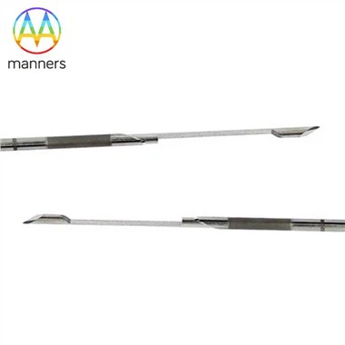
Cutting-Edge Geometry: The "Icebreaker" of Piercing
The shape of the needle tip directly affects the smoothness of penetration and the degree of tissue displacement. Common tip designs include:
- Beveled tip: Similar to a syringe needle, sharp and easy to penetrate, but may push tissue aside rather than cleanly cutting it, suitable for fine-needle aspiration.
- Tru-Cut type: Features an internal sampling groove and an outer sheath with a cutting tube. When the stylet enters the lesion, the outer sheath slides forward, cutting and sealing the tissue protruding into the groove. This design enables retrieval of relatively intact tissue cores and is the classic structure for core needle biopsy.
- Spring-driven: Modern biopsy guns typically use high-speed spring mechanisms, allowing the stylet to complete the "advance-cut-retract" motion in an extremely short time, significantly reducing tissue dragging and tearing, thereby improving sample success rates.
Microscopic Wisdom of the Sampling Slot
The volume, edge sharpness, and surface finish of the sampling groove are equally critical. A longer groove can accommodate more tissue, but if the groove is too shallow, the tissue may become distorted under compression. High-end needle cores feature grooves with laser-polished edges or special coatings to ensure clean, precise cutting, minimizing tissue charring or carbonization. Some newer needle designs also incorporate tiny hooks or textured patterns within the groove to securely hold soft, slippery fat tissue, preventing specimen loss during needle withdrawal.
Association Between Size and Diagnostic Accuracy
Studies show that the diagnostic accuracy of core needle biopsy using a 14G needle can exceed 95%, whereas with an 18G needle, the slightly smaller sample volume increases the risk of misdiagnosis or underestimation of lesion severity. For assessing microcalcifications, vacuum-assisted biopsy with larger-caliber needles (such as 9G) enables collection of sufficient tissue for radiographic confirmation, significantly improving detection rates for ductal carcinoma in situ. Therefore, physicians must carefully balance "obtaining adequate tissue" with "minimizing trauma," and this balance hinges on a deep understanding of needle design parameters.