Straight Tip, Clear Margin
Oct 02, 2026
Patients rarely fear the procedure; they fear the comeback. The fibroid that was "treated" and then regrew from a thin living rim; the follow-up scan that shows a shadow no one explained; the second session no one budgeted for. For the clinician, the parallel anxiety is the viable rim - a 2 mm ring of perfused fibroid left outside the thermal ellipse, quiet for months, then quietly enlarging. The pain is not failure of energy but failure of margin planning. Straight tips help here more than most tools, because their thermal zone is a predictable cylinder - but only if tip exposure, power, and overlap are matched to lesion geometry with intention. Mismatched settings turn the straight tip's predictability into a liability: a too-short exposure leaves the rim; a too-long exposure threatens the serosa.
The mechanism, restated for the margin-minded operator: at ~400 kHz, ionic oscillation generates frictional heat; above ~60°C cell water leaves, proteins coagulate, and the lesion passes into aseptic necrosis. On a straight tip, the lethal isotherm forms a cylinder around the exposed segment, with a softer transition zone at each end. By advancing the needle in 5 mm steps and overlapping half the exposed length, the operator stacks cylinders into a continuous necrotic core that reaches the periphery. The straight geometry turns margin control into arithmetic: no angle correction, no prong spread, just depth steps and overlap fractions.
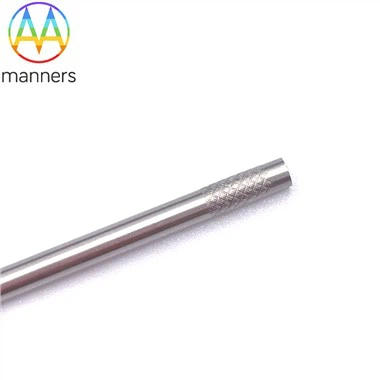
Device choices for margin work are deliberately narrow. A 3 mm exposure suits thin submucosal lesions where the endometrial margin is precious. A 5 mm exposure serves the common 3–4 cm intramural fibroid. A 10 mm exposure is reserved for deep masses with ≥1.5 cm myometrial cover and continuous serosal monitoring. Dual-mark straight tips - two laser rings marking core zone and margin zone - let the operator visualize the intended lethal band on the shaft itself. OEM matched sets, where exposure is fixed per lesion-class protocol, remove the temptation to improvise.
The guide is almost a margin liturgy. Draw the intended non-perfused zone on the ultrasound freeze-frame before energizing. Ablate the core first, then step outward to the rim. After each step, rescan with color Doppler; if a peripheral blush remains, add one more 5 mm track rather than raising power. Resist the urge to declare success when the gray-scale image "looks smaller" - only a non-perfused core on contrast ultrasound counts. Document the overlap fraction and final NPV ratio. Patients are counseled pre-procedure: a residual inactive shadow is expected; symptoms improve before volume disappears.
Programs that have adopted non-perfused-volume (NPV) as the primary endpoint tell a consistent story. Satisfaction scores rise not because fibroids vanish faster, but because expectations are honest. In a series of 3–5 cm intramural fibroids treated with straight-tip moving ablation, most patients reported meaningful menorrhagia relief even when 20–30% of the original volume remained as an inert scar. The straight tip made this reproducible: stacked cylindrical zones, measurable overlap, a margin you can draw on paper before you touch the patient.
Elevating the idea: a clear margin beats a dramatic result. Straight tips turn thermal fibroid therapy from bedside folklore into drawn geometry - a cylinder here, an overlap there, a quiet rim that is planned, not hoped for.
Forward recommendations: teach "isotherm thinking" in every RF course, not just "how to press the pedal." Print the lethal-zone diameter on the pouch for the chosen exposure. Explore ultrasound overlays that project the 60°C isotherm as a faint ring on the live frame. Stop rewarding "fibroid gone" marketing and start rewarding documented NPV at 3 and 6 months. The straight tip's future is not a sharper point - it is a visible, drawable margin.