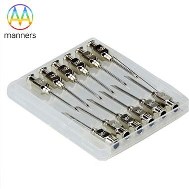
Starting From The Flow Channel Structure Of The Veress Needle
Jun 18, 2026
https://en.wikipedia.org/wiki/Veress_needle
1. Why Does a "Needle" Require a Verification Ritual?
Immediately after inserting the Veress needle, most people's attention jumps straight to "connecting the insufflator, inflating, and inserting the scope." However, experienced surgeons always perform three preliminary steps before connecting the main gas line-steps that exist entirely because of the structural design of the Veress needle.
Recall the gas pathway:
Insufflator → Standard Luer Interface → Stopcock (open) → Inner Stylet Lumen → Side Port of Stylet → Interstice between Outer Cannula and Inner Stylet → Abdominal Cavity.
Along this path, every piece of information available to you is hidden in "how the fluid flows through this slender channel."
2. The Three-Step Verification Method - Each Step Corresponds to a Structural Feature
Step 1: Patency Check - Confirming the Lumen is Not Obstructed
Mandatory pre-procedure: Irrigate the inner lumen with saline. Inject from the hub and observe whether water exits smoothly from the distal side port. Then, occlude the tip and inject forcefully to check if any leakage occurs at the hub or valve seat.
The logic is simple: if the side port is clogged with protein residue from a previous surgery, the gas pathway becomes a "blind-ended tube." CO₂ cannot enter, yet the insufflator-whose pressure sensor is located proximally-assumes everything is normal. You end up with a "non-distending abdomen."
Step 2: Initial Aspiration Test - The Side Port as a Sampling Window
After puncture but before formal insufflation, attach a 5 mL syringe to the hub stopcock and perform gentle aspiration:
Aspirating bowel contents: Indicates the needle is in the bowel lumen. Remove the needle and reinsert at a new site.
Aspirating fresh blood: Suggests entry into the omentum, abdominal wall vessels, or potentially the inferior vena cava (extremely high risk, especially since major vessels lie only 1–2 cm beneath the umbilicus in very thin patients).
No return but negative pressure observed in the syringe: Consistent with the negative pressure characteristics of the peritoneal cavity (a positive sign).
This step is only possible due to the hollow-core and side-port design. If the Veress needle were merely a solid protective rod, you couldn't aspirate anything; you would simply be gambling.
Step 3: Saline Drop Test - A "Free X-ray" of Intra-abdominal Negative Pressure
Classic maneuver: Attach a syringe filled with saline to the needle hub without pushing; let gravity and pressure differentials act.
Saline slowly siphoned in (visible drop in meniscus): Suggests the needle tip is in the free peritoneal cavity.
Saline remains stationary or refluxes: Highly suspicious for a pre-peritoneal location (insufflation zone) or tissue embedding.
Only then connect the CO₂ line and initiate low-flow insufflation (starting at 1 L/min, not 6–10 L/min immediately). Interpret the pressure curve:
|
Pressure Behavior |
Interpretation |
|---|---|
|
Initial negative reading (~ -2 to -4 mmHg), slowly rising to set value |
Likely intraperitoneal ✅ |
|
Pressure spikes rapidly, flow rate near zero |
Tip impacted on tissue/omentum or still pre-peritoneal ⚠️ |
|
Sustained low pressure, abdomen fails to distend |
Gas escaping into wrong space (subcutaneous emphysema / Retzius space) ⚠️ |
3. Structural Defect Turned Diagnostic Advantage - The Duality of the Side Port
The presence of the side port should theoretically be viewed as a design compromise that weakens the cross-section (creating openings in a thin-walled tube inevitably affects bending strength). However, precisely because the hole exists, the Veress needle acquires a triple identity: an insufflation channel + an aspiration window + a negative pressure probe.
Conversely, if you discover during procurement or inspection that a batch of Veress needles has "everted burrs on the side port" or "eccentric hole positioning causing a 50% reduction in effective ventilation area," do not treat it merely as a manufacturing complaint. Realize that this directly impacts the diagnostic credibility during surgery, not just the flow rate metrics.
4. A Clinical Engineering Perspective in One Sentence
The structure of the Veress needle is not just for "getting in"-its hollow core, side port, and stopcock collectively constitute an in-situfluid diagnostic port. Expert surgeons do not rely on "more expensive needles"; they interpret every physical phenomenon within this slender flow channel (negative pressure suction, spontaneous saline flow, the slope of the initial low-pressure rise) as a simplified map of the surgical field.