PTC Needle Application In Biliary Stenosis And Stone Imaging Diagnosis
Nov 11, 2026
1. Clinical Pain Points
Biliary stenosis combined with intrahepatic stones often presents complex anatomical changes. The stenosis may occur at the confluence of left and right hepatic ducts or the common hepatic duct, and the proximal bile duct is dilated with trapped stones. Non-invasive imaging cannot judge the length of stenosis and the patency of downstream duct. ERCP may fail to cross high-grade proximal stricture, leaving clinicians without clear roadmap before treatment. Blind surgical exploration increases bleeding risk and residual stone rate. In addition, mismatched puncture needles easily cause bile leakage, hemobilia and contrast extravasation. Unclear imaging leads to inappropriate treatment plans, such as unnecessary liver resection or insufficient stricture plasty.
2. Working Principle
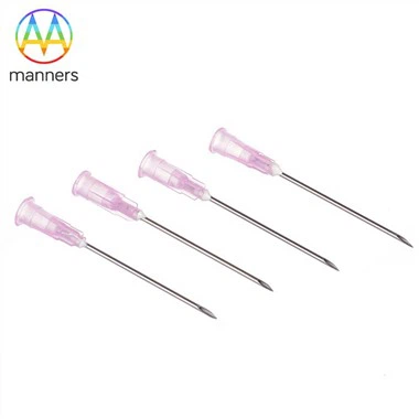
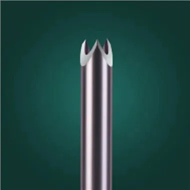
PTC needle delivers contrast medium into the upstream bile duct above stenosis through percutaneous transhepatic access. Under fluoroscopy, the whole biliary tree is displayed, showing the boundary of stenosis, the range of proximal dilation, filling defect shadow of stones and whether the distal duct is patent. The stainless steel needle tube provides stable channel for contrast injection, and the matched stylet protects the lumen during puncture to avoid tissue blockage. The imaging principle relies on the pressure-driven filling of contrast agent, which reflects the elastic property and compliance of bile duct wall. The classic X-ray manifestations captured by PTC include annular stenosis, non-development of isolated duct branches and asymmetric spindle dilation of intrahepatic bile ducts. These signs form the direct basis for judging whether the lesion is stone-related benign stricture or malignant obstructive lesion.
3. Classification of PTC Needles
Diagnostic fine needles (0.25–1.2 mm): designed for patients with mild or non-dilated bile ducts, low incidence of bleeding and bile leakage, suitable for preliminary differential diagnosis. Dual-purpose diagnostic and guide needles (1.2–5 mm): compatible with contrast injection and guidewire passage, used when subsequent PTBD drainage is likely after imaging. Heavy-duty customized needles (>5 mm): applied for thick bile with high viscosity, large-volume bile aspiration and interventional conversion. Surface finish options: polishing reduces tissue friction; electroplating improves wear resistance; ultrasonic cleaning removes micro-particles and machining residues. All needles are ISO9001 certified, customizable from 2D/3D drawings or samples. Packaging can be standard export carton or customized sterile packaging according to hospital requirements.
4. Practical Operation Guide
Preoperative preparation: coagulation function test, routine antibiotic prophylaxis, contrast allergy screening. Mark the target dilated bile duct under ultrasound. Select the puncture path to avoid large intrahepatic vessels and pleural cavity. Ask the patient to hold breath at the moment of needle advancement. After stylet withdrawal, aspirate bile to confirm intraluminal position. Inject contrast slowly under fluoroscopy, stop injection immediately if extravasation appears. Take multiple oblique films to clearly display the stenosis segment at hepatic duct bifurcation. After imaging, withdraw the needle and compress puncture site. Keep patients under observation for 4–6 hours to monitor abdominal pain, fever and hemoglobin change. Record the development status of each hepatic duct branch and the morphological type of dilation.
5. Clinical Experience Sharing
When the imaging shows that part of intrahepatic bile duct fails to develop, operators should not assume it is completely atresia at once. It may be caused by high stone blockage or poor contrast filling. Re-puncture of adjacent duct branches often obtains complete image. For annular stenosis at hepatic duct bifurcation, PTC can measure the stenosis length accurately, which guides the surgical design of duct reconstruction. For patients with biliary cirrhosis, fine-gauge needle is preferred, because cirrhotic liver parenchyma has poor self-healing ability and bleeding risk rises sharply. Operators should control injection pressure, especially for ducts with thin, inflamed walls. Many cases show that PTC provides clearer anatomical roadmap than CT reconstruction for complex bilateral intrahepatic stone disease.
6. Summary
PTC needle is the core consumable to obtain direct cholangiography images for biliary stenosis and stone disease. It solves the problem that non-invasive imaging cannot evaluate the real duct patency above stricture. Different needle specifications match different clinical scenarios from simple diagnosis to interventional transition. Standard puncture and contrast injection protocols reduce major adverse events. PTC imaging signs are the key evidence for distinguishing benign stone stricture from malignant obstruction.
7. Industry Prospect and Suggestion
Future iteration direction focuses on ultrasonic visible needle tip and graduated depth marking to reduce fluoroscopy time. Manufacturers can develop color-coded series by diameter for quick identification in operation room. Clinical departments should build PTC image database to summarize the X-ray characteristics of stationary intrahepatic stones. For patients with high suspicion of biliary malignancy, PTC can be combined with CT for comprehensive evaluation. Medical device suppliers need to maintain stable precision and surface quality while supporting personalized drawing customization, to meet the demand of complex biliary intervention centers worldwide.
< img alt="news-1-1" src="/uploads/24240/news/202604241539186d0ff.png " />