How PTC Needle Resolves Misdiagnosis Of Intrahepatic Cholangiolithiasis
Nov 11, 2026
1. Clinical Pain Points
Intrahepatic cholangiolithiasis in stationary phase often lacks obvious fever and acute abdominal pain. Many patients only show vague upper abdominal discomfort, fatigue and abnormal liver function indicators. Such manifestations are highly similar to viral hepatitis, gastritis and functional dyspepsia, leading to high misdiagnosis rate in primary diagnosis. B-ultrasound has limited resolution for tiny intrahepatic duct stones and subtle duct stricture; CT cannot dynamically display the whole bile duct tree. Without intuitive cholangiography images, clinicians cannot judge whether the stenosis is annular, local or segmental, nor confirm whether spindle-shaped or dumbbell-shaped dilation exists. Repeated misdiagnosis delays intervention, and long-term stone irritation may progress to biliary cirrhosis or malignant transformation. Meanwhile, old thick puncture needles bring high bleeding and bile leakage risks, which restrict the popularization of PTC examination for low-symptom patients.
2. Core Working Principle of PTC Needle for Cholangiography
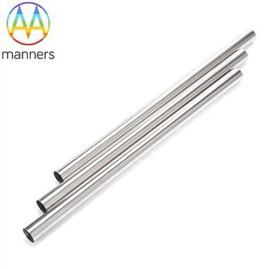
PTC needle is a percutaneous transhepatic puncture and injection instrument made of medical stainless steel. Under ultrasound or fluoroscopy guidance, the needle penetrates liver parenchyma into target intrahepatic bile duct. After bile aspiration confirms correct lumen position, radiopaque contrast medium is injected slowly. The contrast agent fills the bile duct tree, and X-ray captures the morphological features of hepatic duct bifurcation, duct wall, stenosis, dilation and filling defects. Its core principle is to convert invisible biliary anatomical lesions into visible radiographic signs. The needle's smooth inner channel ensures stable contrast injection without air bubble interference, and precise tip design reduces tearing of bile duct mucosa and hepatic vascular injury. It directly visualizes the X-ray features described in clinical guidelines: annular stricture at common hepatic duct or bifurcation, non-development of local intrahepatic ducts, asymmetric duct dilation of liver lobes.
3. Classification of PTC Needles for Cholangiography
By diameter, thin-gauge fine needles range from 0.25 mm to 1.2 mm, mainly used for diagnostic cholangiography on non-dilated bile ducts, with low bleeding risk. Medium-size needles between 1.2 mm and 5 mm balance puncture stability and contrast delivery efficiency, suitable for patients with moderately dilated intrahepatic ducts and suspected stationary stones. Large customized needles above 5 mm are reserved for combined bile aspiration and guidewire exchange, preparing for subsequent drainage surgery. By surface treatment, polished needles have smooth outer surface to reduce liver tissue dragging; electroplated and plated needles improve corrosion resistance; ultrasonic cleaned needles eliminate residual machining oil and debris to meet biocompatibility requirements. Customized models can be produced according to 2D drawings, 3D files or physical samples, including special tip taper, side hole structure and customized length. All products are ISO 9001 certified, packaged in standard cartons or customer-specified packaging.
4. Standard Operation Guide
Before operation, review B-ultrasound and CT data to mark target bile duct, measure puncture depth and angle. Routine prophylactic antibiotics are administrated to reduce cholangitis risk. Place patient in supine or slightly oblique position, select midaxillary line puncture point below the 10th rib. Under real-time ultrasound guidance, advance the PTC needle during breath hold. After removing the stylet, connect syringe and aspirate bile to verify duct access. Inject contrast medium slowly under fluoroscopy control, avoid overpressure injection which causes contrast extravasation. Capture spot films in multiple projection angles to observe left and right hepatic duct development, stricture range and stone shadow. After imaging is completed, withdraw the needle gently, apply local compression and monitor vital signs for bleeding, biliary peritonitis and fever. Record whether the imaging shows asymmetric, spindle-shaped or dumbbell-shaped dilation of intrahepatic bile ducts.
5. Practical Clinical Experience
For stationary-phase intrahepatic cholangiolithiasis without infection, operators should prefer thin-gauge PTC needles to minimize trauma. Do not inject contrast agent blindly if no bile returns after puncture, to prevent hepatic parenchyma contrast retention. When part of intrahepatic duct fails to develop, adjust puncture site instead of increasing injection pressure. For patients combined with biliary cirrhosis, reduce contrast dosage and injection speed because fragile bile duct walls are prone to rupture. Many clinical cases prove that PTC imaging can correct the previous misdiagnosis of hepatitis or gastritis. Even if B-ultrasound shows only mild echo change, PTC can expose hidden annular stenosis and proximal stone shadow. Operators need to distinguish benign stricture from malignant stenosis by duct edge regularity, combined with CT findings.
6. Summary
PTC needle for cholangiography fills the gap between non-invasive imaging and surgical exploration. It solves the core pain point of high misdiagnosis rate of stationary intrahepatic cholangiolithiasis. As a precision injection and puncture device, it realizes direct visualization of biliary tree morphology. Rational selection of needle diameter and surface treatment scheme can match different biliary dilation degrees and patient conditions. Standardized puncture and contrast injection procedures effectively control major complications.
7. Prospect and Suggestion
In the future, integrated ultrasound-visible PTC needles will further improve puncture accuracy and reduce fluoroscopy radiation exposure. Manufacturers can expand standard size series while maintaining flexible customized service for special clinical requirements. Hospitals should organize regular training on PTC image reading, focusing on identifying classic X-ray signs of intrahepatic stones. For patients with unexplained liver function abnormality and upper abdominal pain, PTC examination should be included in differential diagnosis workflow after excluding gastritis and hepatitis. Joint evaluation of PTC and CT will improve early detection rate of biliary malignant lesions in cirrhotic patients.
< img alt="news-1-1" src="/uploads/24240/news/202604241539186d0ff.png " />