Operation Of Liver Biopsy
Nov 10, 2022
1. Preoperative preparation, puncture method and ptc needle angiography.
2. ptc angiography was performed with a 22-gauge fine needle to determine the location and nature of the lesion.
3. According to the angiographic results, a thick, straight and horizontal bile duct was selected for internal drainage intubation.
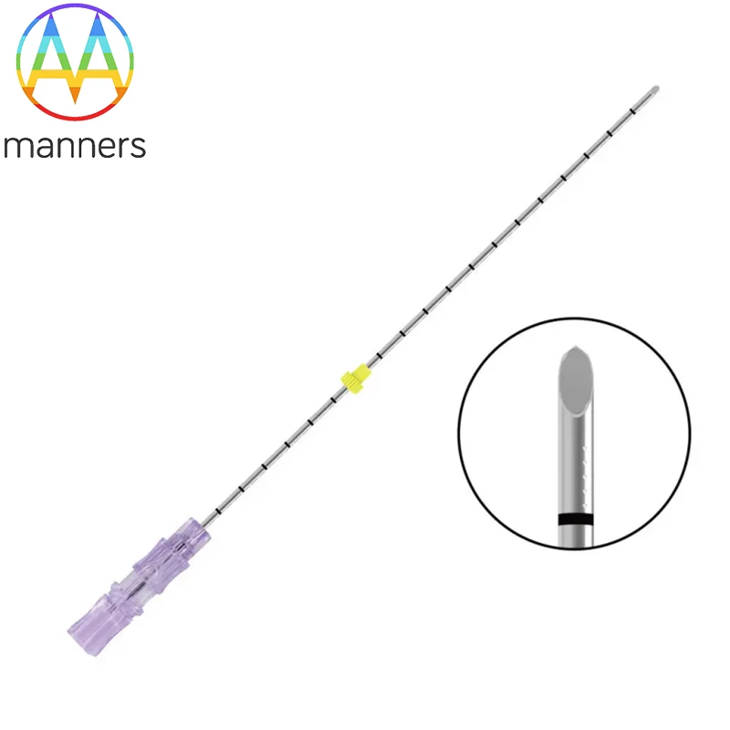
4. Make another puncture point from the 8th rib of the right midaxillary line, and poke a small incision on the skin with a sharp knife after local anesthesia. The patient is instructed to stop breathing, and the thick needle is quickly inserted into the pre-selected bile duct under the surveillance of TV. When there is a sense of breakthrough into the bile duct, the needle core is pulled out, and the guide wire is inserted into the distal bile duct or duodenum through the obstructed end or narrow segment, and the direction is constantly rotated and changed to make the guide wire enter the distal bile duct or duodenum through the obstructed end or narrow segment, exit the puncture needle, and expand the channel with the dilator tube. The multi-lateral hole catheter was passed through the obstruction end or the stenosis segment with the guide wire, so that the side hole of the catheter was located above and below the obstruction end or the stenosis segment, and the catheter was fixed. After the bile was smoothly discharged from the catheter, the contrast agent was injected and the film was taken.
5. A week after drainage, angiography was performed to observe the catheter position and drainage effect.