Needle For Intraosseous Access In Cardiac Arrest
Nov 20, 2026
The Ten-Second Window Inside a Two-Minute Cycle
Cardiac arrest compresses every clinical decision into a two-minute rhythm cycle. Within that cycle, compressions must continue, the airway must be managed, the rhythm must be interpreted, and drugs must be delivered. Access competes with all of them for the same pair of hands. This is the context in which a needle for intraosseous access proves its value: it is the only vascular route that can be established inside the compression interval without interrupting chest compressions.
The arithmetic is unforgiving. A peripheral attempt in a pulseless patient means palpating for a vein that has no pressure behind it, often while the patient is being moved or while a mechanical compression device occupies the groin and neck. Two failed attempts can consume the better part of a cycle. Drug administration then slips to the next rhythm check, and the next, until epinephrine intervals stretch well beyond what the protocol intends. The delay is rarely recorded as an access delay; it appears in the record as a slow drug time.
Setting a hard threshold resolves the ambiguity. If peripheral access is not achieved quickly in a patient with no palpable pulse, the intraosseous route should be chosen rather than pursued as a last resort. The decision is not an admission of failure; it is a scheduling choice that protects the one resource the patient cannot spare.
Why Marrow Perfusion Survives Cardiac Standstill
Peripheral veins collapse in arrest because they are low-pressure conduits dependent on forward flow. The medullary sinusoids behave differently. They are enclosed within a rigid trabecular scaffold, and they communicate directly with the central venous system through emissary veins. Even when cardiac output ceases, that anatomical relationship persists, and fluid instilled into the cavity reaches the central circulation as soon as return is re-established, and continues to do so during effective chest compressions.
Drug onset through this route is comparable to intravenous administration, which is why resuscitation guidelines treat the two routes as equivalent for arrest medications. The practical corollary is that dose adjustment is unnecessary. Epinephrine, amiodarone, lidocaine, bicarbonate in specific indications, and volume expanders are given at standard intravenous doses, followed by a flush that is large enough to move the drug out of the medullary channel and into the central compartment.
That flush is where many administrations quietly fail. A small flush leaves drug resident in the cavity, and the resulting clinical non-response is misinterpreted as a drug failure rather than a delivery failure. A deliberate flush of at least ten milliliters after each drug, with the limb elevated where feasible, converts a nominal administration into a real one.
Device Classes and the Variables That Matter in Arrest
Three device families are used in resuscitation. Manual needles demand sustained axial force and a twisting action, which is difficult while wearing gloves in a moving vehicle. Spring-loaded impact devices deliver a single fixed stroke and are fast, but offer limited control in fragile bone. Powered drivers convert stored energy into rotation or oscillation, and when fitted with a cutting-tip needle they open the cortex by machining a channel instead of splitting it.
In arrest, the variables that matter are speed, stability, and independence from operator strength. A cutting geometry driven by a low-power motor allows a clinician to seat the device in as little as ten seconds without applying body weight, which matters when the patient is on a narrow stretcher, when the operator is kneeling, or when a second clinician is performing compressions centimeters away. The resulting needle sits rigidly enough to survive transport and to accept pressure-assisted infusion without dislodging.
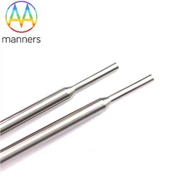
Needle specification follows the same logic as any other setting. A 15G stainless steel needle covers most adult patients, with custom lengths and diameters available for extremes of body habitus or for pediatric use. Because these are injection and puncture instruments rather than commodity consumables, suppliers commonly produce them to 2D or 3D drawings or against a submitted sample, which allows a service to standardize one needle geometry across adult and pediatric kits and to receive consistent stock in standard cartons.
Insertion Sequence During Ongoing Compressions
The sequence should be rehearsed until it requires no discussion. Assign the procedure to a single clinician and state the assignment aloud; compressions do not pause for access. Choose the proximal tibia on the flat medial surface, approximately two centimeters below the tibial tuberosity. It is the least disruptive site during compressions because it is far from the airway, does not interfere with defibrillator pad placement, and remains accessible when a mechanical compression device is in use. The proximal humerus is the alternative when the lower limbs are trapped or when the tibial cortex is unusable.
Cleanse the site and allow the antiseptic to dry while the next rhythm analysis proceeds. Stabilize the leg against the stretcher rather than against a moving surface. Seat the needle with the driver until resistance falls away, then remove the driver while holding the hub steady. Confirm by aspirating marrow if possible, flushing against resistance, and checking that the needle stands unsupported. Apply the stabilization dressing, then give the first flush in a pulsed manner to clear debris from the channel.
Two details separate a routine insertion from a complicated one. First, do not release the needle before the dressing is applied; an unsecured device is easily levered out during the next transfer. Second, record the insertion time on the same clock used for drug intervals so that the access timestamp and the pharmacology timeline share a reference.
Field Lessons and the Errors That Break the Cycle
The most common error is delaying the decision. Crews often begin a peripheral attempt because it is familiar, then persist because abandoning it feels like conceding. The cost is measured in skipped drug cycles, not in the failed puncture itself.
The second error is site selection driven by convenience rather than by the resuscitation. A femoral or humeral attempt that requires repositioning the patient can cost more time than it saves, and an attempt in a limb that is being manipulated by another rescuer invites dislodgement. The third error is inadequate flushing, described above, which produces a pharmacologically invisible dose. The fourth is failing to reassess the site after each patient movement; extravasation in arrest is easy to miss because swelling competes with edema from resuscitation.
A fifth error is conceptual: treating the intraosseous line as a permanent solution. It is a bridge. Once return of spontaneous circulation is achieved and the patient reaches an environment where reliable intravenous or central access can be obtained, the device should be exchanged. Keeping a bridging needle in place because it is working is a reasonable short-term choice and a poor long-term one.
Rethinking Access as a Rhythm Intervention
Stop describing vascular access as preparation and start describing it as part of the rhythm. Every intervention in arrest is judged by whether it fits inside the cycle without degrading compressions. By that measure, an access route established in ten seconds is not an accessory to resuscitation; it is a component of it.
The reframing changes preparation. Equipment is laid out before the call rather than assembled during it, the access clinician is named at the start rather than recruited midway, and the threshold for switching routes is agreed in advance. None of that requires new technology; it requires treating the device already in the bag as a first-line instrument.
Design Trends and Procurement Questions
Development in this category is converging on three themes: gentler energy delivery, geometry that produces a clean cortical channel, and single-use kits that eliminate assembly. Cutting-tip designs paired with low-power drivers embody all three, because they reduce the force an operator must generate and therefore reduce the variability between a strong clinician and a tired one.
Four specification questions are worth asking. Does one driver cover adults, children, and high-BMI patients? Is the tip a true cutting geometry, with energy low enough that insertion does not depend on operator weight? Can the manufacturer supply to your 2D or 3D drawings or to a physical sample, and hold that geometry across orders? And does standard carton packaging suit vehicle restocking without repacking? A device that answers all four becomes standard equipment rather than a specialist item, and standard equipment is what gets used at two in the morning.