Pediatric Intraosseous Access Without Force
Nov 20, 2026
Why a Child's Cortex Changes the Whole Technique
The same procedure that is forgiving in an adult is unforgiving in a child. A child's cortical bone is thinner, more elastic, and far easier to traverse than the dense cortex of a middle-aged adult. That sounds like good news for the operator, and it is, until the needle passes through the far cortex and into the soft tissues behind it. Over-penetration is the characteristic complication of pediatric intraosseous insertion, and it is caused less by inexperience than by technique borrowed from adult practice.
The margin for error is small in both directions. A needle that stops short produces no flow and a false confirmation. A needle that travels too far produces extravasation into the subcutaneous space or, worse, into the subperiosteal compartment, with compartment syndrome as a downstream risk. Meanwhile the anatomy imposes constraints: the medullary cavity in an infant is narrow, the epiphyseal plate must be avoided, and the landmark distances used in adults must be scaled down rather than copied.
The practical conclusion is that pediatric access demands a method in which depth is controlled by the device and the operator's judgment, not by how hard the operator pushes. Any technique that rewards force is a technique that punishes children.
The Mechanics of Controlled Penetration
Insertion into bone fails in one of two ways: the needle does not enter, or it enters too far. Manual needles address the first problem with force and the second with the operator's reaction time, which is why they are difficult to use well in small bone. A rotating or oscillating cutting tip addresses both problems differently. The tip machines a channel through the cortex, so forward progress requires very little axial load, and the transition from cortex to marrow produces a distinct change in resistance that the operator can feel and respond to immediately.
Pairing that tip with a low-power driver completes the logic. Because the motor supplies the cutting energy, the clinician does not lean on the device, and therefore does not convert body weight into penetration depth. In a ten-second insertion, the operator's role shifts from applying pressure to holding the device steady and watching for the end point. That is a meaningfully safer division of labor when the patient weighs twelve kilograms.
The same principle explains why tip geometry is not interchangeable across age groups. A wedging tip requires axial force proportional to bone density, which means the force that is appropriate for an adult is excessive for a toddler. A cutting geometry scaled to the needle diameter keeps the required force low across the whole range, which is what allows one technique to serve both ends of the patient spectrum.
Matching Needle Length and Gauge to Small Anatomy
Gauge and length are separate decisions that are often conflated. Gauge governs flow and resistance; length governs reach. For most pediatric patients a 15G needle provides adequate flow for resuscitation volumes while keeping the outer diameter modest. Length is where pediatric practice diverges: a needle chosen for an adult tibia will reach the far cortex in a small child.
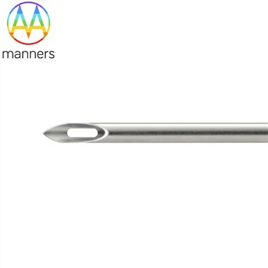
The safest approach is a system built around a needle for intraosseous access that offers multiple lengths behind a single driver, so that length is a deliberate choice rather than a default. Needles are typically stainless steel to retain a sharp edge and resist bending, and manufacturers commonly produce to custom specifications from 2D or 3D drawings or from a supplied sample. That flexibility lets a service order a shortened pediatric variant matched to its adult geometry, delivered in standard cartons with consistent labeling.
Landmark selection is equally size-dependent. In older children the proximal tibia, on the flat medial surface roughly one to two centimeters below the tibial tuberosity, is standard. In infants and very young children, the same site is used with reduced distances, and alternative sites such as the distal tibia or distal femur are chosen according to device labeling and local protocol. Whatever the site, the epiphyseal plate is avoided, and the needle is directed away from it.
A Step-by-Step Approach for Infants and Children
Begin by confirming that peripheral access has failed or will consume unacceptable time, and that the child's condition warrants immediate vascular access. Assign one clinician to the procedure and one to hold the limb firmly; a moving leg is the most common cause of a badly placed needle in a small patient. Expose the site fully and identify the tibial tuberosity by palpation rather than by eye, since soft tissue obscures landmarks in infants.
Cleanse the skin and allow it to dry. Hold the leg against a firm surface, not against a rescuer's moving hand. Seat the needle with the driver using light, steady control, and stop the moment resistance disappears; do not continue for a fixed count. Remove the driver while stabilizing the hub, then confirm placement by gentle aspiration, a small flush against resistance, and observation of a needle that stands without support. Apply the stabilization dressing before releasing the device.
Two pediatric-specific steps follow. First, secure the limb against the stretcher with the dressing and, where the device design allows, a length of tape that prevents flexion at the knee or hip. Second, reassess the site after every transfer, because a needle that is adequate in a resuscitation bay can migrate during a move to the ambulance. In conscious children, local anesthetic infiltration before insertion, and slow lidocaine infusion after confirmation, reduces the pain of the flush without delaying access.
Field Experience and the Errors That Injure Children
The most damaging error is applying adult force to a pediatric cortex. It presents as a needle that is fully seated, flushes with little resistance, and then produces swelling within minutes. The second error is choosing length by habit. A crew that stocks one needle length for all patients will eventually over-penetrate a small child, and the failure will look like a device problem rather than a selection problem.
The third error is inadequate immobilization. In infants, the medullary cavity is short, and a few degrees of limb movement during insertion changes the trajectory enough to matter. The fourth is treating a successful flush as complete confirmation. Free flow can occur with the tip in soft tissue if the tissue is loose; the standing needle, the absence of swelling, and a clinical response to the first drug are the more reliable signs.
A fifth error is the assumption that children require a different, more cautious drug strategy once access is obtained. They do not. Doses are weight-based, as they are intravenously, and the route does not alter the pharmacology. Over-caution here produces under-dosing, which is its own form of harm.
What Gentle Access Protects
The value of a force-free technique is not comfort for the clinician. It is the preservation of structures that cannot be inspected after the fact: the far cortex, the periosteum, the epiphyseal plate, and the soft tissue compartments of a small limb. Once those are violated, the consequences are not visible on the monitor, and they may not become apparent until the child is on a ward hours later.
Gentle access also protects the resuscitation itself. A technique that does not require strength can be performed by any trained team member, in any posture, on any surface. That widens the set of people who can solve the access problem in the first minute.
Design Directions and Selection Criteria
Developments in this category point toward finer depth control and simpler kits. Cutting-tip geometries with low-power drivers are becoming the default because they decouple penetration from operator strength. Color-coded length options, integrated depth referencing, and pre-assembled stabilization dressings reduce the number of decisions made under pressure.
For teams specifying equipment, the practical test is whether one platform can serve a neonate and a large adolescent without changing technique. Ask whether the supplier can provide a shortened variant to your 2D or 3D drawings or to a sample, whether the needles are stainless steel with a genuine cutting tip, whether the driver's energy is low enough that insertion does not depend on operator weight, and whether standard carton packaging supports separate pediatric stocking. A needle for intraosseous access that meets these criteria stops being a compromise choice for children and becomes the routine first-line route when peripheral access fails.