How Histological Biopsy Needle PTC Optimizes Specimen Quality In Cervical Pathology Diagnosis
Nov 11, 2026
1. Clinical Pain Points
Poor specimen quality is a frequent cause of pathology reporting failure. Blunt biopsy needles crush epithelial layers, destroying the layered structure of cervical tissue. Insufficient tissue volume makes it impossible for pathologists to judge whether dysplasia extends into the stroma. When samples are fragmented, laboratories often issue "non-diagnostic" reports, forcing patients to return for a second biopsy and increasing psychological burden. Many clinics use universal biopsy instruments for all cervical cases, without differentiating superficial erosion from deep endocervical lesions. CTC cytology can only offer cell-level clues, and the diagnostic value collapses if the subsequent biopsy specimen is unqualified. In addition, residual burrs on needle surfaces may cause extra mucosal laceration and prolonged post-procedure bleeding.
2. Working Principle
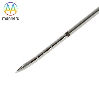
Histological biopsy needle PTC obtains intact cervical tissue blocks by controlled cutting. Its sharp, precisely machined tip separates target epithelium and stroma cleanly without squeezing cells. The hollow lumen locks the tissue sample during withdrawal to prevent shedding. Colposcopy magnifies the cervix 40 times to pinpoint erosion and suspicious areas, guiding the needle to sample the highest-risk region. The harvested tissue is fixed and sectioned for histology, which is the definitive diagnostic method. Surface treatment determines friction performance: smooth surfaces reduce tearing, while clean surfaces eliminate foreign-body contamination that may interfere with microscopic observation. The needle's mechanical stability ensures consistent cutting force, so specimens maintain natural histological architecture for accurate grading of CIN lesions or invasive cancer.
3. Classification of Histological Biopsy Needle PTC
Fine sampling needles (0.25–1.2 mm): for superficial squamous epithelium of mild erosion, preserve intact epithelial layers, low bleeding risk. Standard diagnostic needles (1.2–5 mm): mainstream choice for most suspicious lesions, delivering enough stromal tissue for complete pathological evaluation. Custom deep-sampling needles (5–30 mm): for endocervical canal lesions and suspected glandular lesions, customizable length and tip angle. Surface finish categories: polishing for low friction and clean cuts; electroplating for wear resistance during repeated sampling; ultrasonic cleaning to remove micro-particles and machining residues before packaging. All products are ISO9001 certified, customizable from 2D/3D drawings or samples, with flexible packaging options.
4. Practical Operation Guide for High-Quality Specimen Acquisition
Preoperative preparation: confirm colposcopic target areas and CTC screening results. Select needle size according to lesion depth. Inspect the needle tip visually before use to exclude burrs or deformation. Under colposcopy, apply acetic acid and iodine to mark abnormal zones. Stabilize the cervix gently, align the needle perpendicular to the lesion surface, execute one rapid steady sampling motion and avoid twisting. Immediately transfer the whole specimen into fixative without squeezing. If multiple lesions exist, label each sample with its anatomical position. After sampling, apply local compression and observe bleeding. Record whether the specimen is intact, and feed back poor sampling results to the device supplier.
5. Clinical Experience Sharing
The biggest enemy of high-quality biopsy samples is slow, wobbly needle movement. A sharp polished PTC needle completes cutting in one action, which greatly reduces crush artifact. Ultrasonic cleaning is essential; tiny metal particles left on the needle may appear as artifacts under the microscope and confuse pathologists. Medium-size needles work best for transformation zone lesions, which are the highest-risk region for cervical precancerous changes. Operators should not choose oversized needles simply to get "more tissue", because excessive depth may sample normal deep tissue and miss the surface dysplastic epithelium. When CTC shows atypical glandular cells, customized angled PTC needles targeting the endocervix are preferred, as surface sampling often yields false-negative results.
6. Summary
Specimen integrity directly decides whether biopsy can fulfill its role as the definitive diagnostic test after inconclusive CTC screening. Histological biopsy needle PTC's tip geometry, material and surface treatment collectively determine specimen quality. Classified needle selection matching lesion depth minimizes crush damage and sampling failure. Standardized sampling techniques preserve tissue architecture for reliable histological grading.
7. Industry Prospect and Suggestion
Manufacturers should add automated tip sharpness inspection in production lines to reduce defective batches. They can launch a specimen-oriented selection table for gynecologists. Future iterations can integrate anti-adhesion coating to stop tissue sticking inside the needle lumen. Clinical departments should establish communication channels between colposcopists and pathologists to collect feedback on biopsy needle performance. High-quality PTC biopsy needles will lower the rate of non-diagnostic pathology reports and improve the efficiency of cervical cancer screening chains.
< img alt="news-1-1" src="/uploads/24240/news/202604241539186d0ff.png " />