Historical Review: The Evolution Of Meniscus Repair Techniques
Apr 15, 2026
Historical Review: The Evolution of Meniscus Repair Techniques
From the "era of total resection" to "precision repair," how has the philosophy of meniscus treatment evolved over time? And what revolutions in medical understanding lie behind each technological breakthrough?
Stage One: The Era of Resection (Pre-1980s)
Before the 1980s, the dominant approach to meniscus injuries was total resection. The prevailing belief was that the meniscus was merely an embryonic remnant with no significant function and should therefore be removed when injured. This thinking led to a large number of total meniscectomies. While patients often experienced short-term pain relief, the long-term consequences were severe - a sharp rise in osteoarthritis incidence and a marked reduction in the age at which knee replacements became necessary.
A key figure of this era was British surgeon Fairbank, who in 1948 first systematically described radiographic changes following meniscectomy, including joint space narrowing, osteophyte formation, and flattening of the femoral condyles - later known as Fairbank's triad. Although he warned of the serious repercussions of meniscus removal, resection remained the mainstream choice due to a limited understanding of meniscal function.
Stage Two: Partial Resection and Suturing Exploration (1980s–1990s)
With the popularization of arthroscopic technology and advances in biomechanical research, the medical community gradually recognized the meniscus's vital roles: load distribution, improving joint congruity, and providing stability. In 1982, Henning reported the first arthroscopic meniscus suturing technique, marking the beginning of the meniscus preservation era.
A major breakthrough came from Cooper and colleagues' in-depth study of meniscal blood supply. They divided the meniscus into the red zone(well-vascularized), red-white zone(border area), and white zone(avascular), providing a theoretical basis for selective resection. However, the posterior root of the medial meniscus remained a "no-stitch zone" due to its difficult location, poor exposure, and limited healing capacity.
Stage Three: Difficult Exploration of Root Repair (Early 2000s–2010)
In the early 21st century, advances in MRI technology allowed the medical field to recognize the prevalence and severity of root injuries. In 2006, LaPrade's team provided the first systematic description of the biomechanical consequences of medial meniscus posterior root tears, finding that the increase in joint contact pressure was comparable to that seen after total meniscectomy.
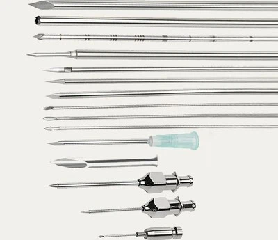
Repair techniques of this period focused mainly on transtibial tunnel pullout methods. Surgeons drilled tibial tunnels and pulled the meniscus root out to fix it to the bone surface. While theoretically viable, two major drawbacks emerged: the "bungee effect" - micro-motion of sutures inside the tunnel causing loosening; and "meniscus cutting" - sutures slicing through fragile meniscus tissue under load.
Stage Four: The Anchor Fixation Era (2010–2020)
With the evolution of suture anchor technology, surgeons began attempting direct fixation of the meniscus root using anchors. This avoided the need for bone tunnel creation, offering a theoretically simpler and less invasive option. However, in practice, conventional anchors had limited insertion angles in the posteromedial compartment, insufficient fixation strength, and a persistently high risk of cutting.
A key development in this stage was deeper biomechanical insight. Numerous studies compared initial fixation strength, displacement after cyclic loading, and failure modes across different repair techniques, providing quantitative evidence for technical refinement. Yet, even up to 2020, clinical outcomes for posterior root tears remained suboptimal, with re-tear rates fluctuating between 20% and 40%.
Stage Five: The Revolutionary Breakthrough of Inverted Anchors (2020–Present)
Professor Han Changxu's team built their innovation upon this historical trajectory. Reviewing the entire developmental history of meniscus repair, they realized that every previous breakthrough had stemmed from a fundamental reconsideration of biomechanical principles. The inverted anchor technique's core innovation lies not in minor tweaks to existing methods, but in a complete reconstruction of repair philosophy.
The historical significance of this technique is that it achieves, for the first time, an optimal balance between biomechanics and clinical feasibility. Through inverted anchor implantation, stress distribution at the repair site becomes more uniform, eliminating stress concentration. Special angular design ensures that suture force aligns with the meniscus's physiological load direction, minimizing abnormal shear forces.
The advancement of history always follows a spiral path. From total resection to partial resection, from attempted suturing to precision repair, each step represents a deepening of medical understanding. The inverted anchor technique may not be the final destination of meniscus repair, but it stands as a significant milestone in the present stage - paving the way for even more advanced solutions in the future.
If you want, I can also create a chronological table summarizing these five stages with timelines, key technologies, and limitations for a clearer presentation. Would you like me to prepare that?