Historical Evolution And Technical Principles Of Intraosseous Access Needles
May 10, 2026
Introduction: From Battlefield First Aid to a Pillar of Modern Emergency Care Systems
Intraosseous (IO) access needles are breakthrough emergency medical devices. By establishing access through bone (typically long bones), they directly connect to the abundant vascular network within the medullary cavity, enabling rapid and reliable infusion of fluids and medications. Though the technology dates back to the early 20th century, its true revival and standardized clinical application have taken shape over the past three decades, driven by conceptual innovations in emergency medicine.
The medullary cavity is a unique physiological space filled with numerous non-collapsible venous sinuses, which connect to the systemic circulation via nutrient veins and emissary veins. During circulatory failure, peripheral blood vessels may collapse completely; by contrast, the vascular structures inside the medullary cavity remain patent due to being enclosed by rigid bony tissue, making them the only reliable vascular access at such critical moments. This anatomical characteristic forms the physiological foundation of intraosseous access technology.
Technical Principle: The Medullary Cavity as a Non-Collapsible Venous Pathway
The core principle of intraosseous access lies in the fact that the bone medullary cavity is essentially a rigid, incompressible chamber rich in vascular structures. Even in severe hypovolemic shock or cardiac arrest, when peripheral veins collapse completely and central venous access cannot be established rapidly, the venous sinuses within the medullary cavity stay open. Protected by hard cortical bone, they do not collapse under external pressure like soft-tissue veins.
Medullary blood circulation connects to the systemic circulation, supplied primarily by nutrient arteries and drained through nutrient and emissary veins. Once medications or fluids are infused into the medullary cavity, they are quickly absorbed into these venous sinuses and enter the systemic venous circulation via nutrient veins. Studies show that fluids administered via the intraosseous route reach the central circulation in an average of 10 to 30 seconds - comparable to central venous access and far faster than most peripheral venous routes.
Device Evolution: Revolution from Manual to Semi-Automatic Design
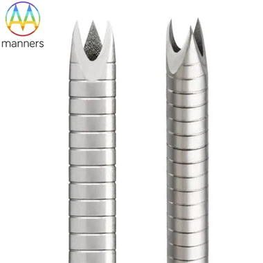
Early intraosseous needles (such as the Jamshidi needle) were essentially modified bone marrow biopsy needles. They required substantial force and technical skill to penetrate the bone cortex, with limited success rates. In the 1980s, as emergency medicine emphasized the golden hour concept, intraosseous technology underwent its first innovation: purpose-built IO needles emerged with sharper tips and higher structural strength, though operation remained fully manual.
The real revolution came in the early 21st century with the invention of semi-automatic and automatic IO puncture devices. Powered by springs or batteries, these devices drive specialized puncture needles into bone with controlled force and speed, greatly lowering technical barriers and saving procedural time. Representative products include:
EZ-IO System: Battery-powered with a specially designed needle tip, capable of accessing multiple sites including the adult tibia, humerus, and sternum; puncture completion usually takes less than 30 seconds.
FAST1 Sternal Puncture Device: Designed specifically for sternal access, featuring a stable platform and depth precision control to avoid injury to posterior mediastinal structures.
BIG (Bone Injection Gun): Originally developed for military use with spring-driven operation, now available in multiple upgraded generations.
These devices have raised the overall puncture success rate from approximately 70% with manual methods to over 95%, while cutting procedural time from several minutes to mere seconds - perfectly meeting the time-critical demands of emergency care.
Anatomical Selection of Puncture Sites
Selecting an appropriate puncture site is critical to successful and safe IO access. Each site carries specific indications and precautions:
Proximal Tibia: The most commonly used site, located 1–3 cm inferior and medial to the tibial tuberosity on the flat medial surface. The cortical bone here is relatively thin with no vital underlying structures, suitable for both children and adults, and the first choice for most emergency scenarios.
Humeral Head: Located 2–3 cm below the acromion and inferior to the greater tubercle of the humerus. Particularly ideal during cardiopulmonary resuscitation, as it does not interfere with chest compressions; care must be taken to avoid injuring the radial and axillary nerves.
Distal Femur: Located a palm's breadth above the superior patellar border on the medial femur. Indicated for pelvic fractures or lower limb trauma where tibial access is contraindicated.
Sternum: Punctured through the sternal body using dedicated devices such as the FAST1 system. It lies closest to the central circulation for the fastest drug onset, yet requires higher technical proficiency and carries a slightly elevated risk of complications.
Distal Radius / Ulna: Mainly used in pediatric patients and rarely in adults.
Milestones in Technical Standardization
The widespread adoption of intraosseous access has been driven by the establishment of authoritative guidelines and standards:
In 2000, the American Heart Association (AHA) first recommended IO access as an alternative to venous access in its Advanced Cardiac Life Support (ACLS) guidelines.
In 2005, the International Liaison Committee on Resuscitation (ILCOR) formally recognized IO access as a first-line method for establishing vascular access during cardiac arrest.
The 2010 AHA guidelines further clarified that intraosseous access should be initiated immediately if venous access cannot be established within 90 seconds in emergency situations.
In 2015, the American College of Surgeons Committee on Trauma incorporated IO access as a standard component of Advanced Trauma Life Support (ATLS) protocols for trauma resuscitation.
Future Outlook: Intelligence and System Integration
Next-generation IO devices are evolving toward greater intelligence and integrated functionality:
Real-Time Feedback Systems: Equipped with pressure sensors and impedance measurement technology to verify needle tip position during puncture, preventing over-penetration or insufficient depth.
Integrated Ultrasound Guidance: Portable ultrasound seamlessly integrated with IO devices to visualize the puncture process and improve first-pass success rates, especially in obese patients or those with anatomical variations.
Intelligent Infusion Flow Control: Automatically adjusts infusion rates based on medullary cavity pressure and patient hemodynamic status to reduce complication risks.
Wireless Monitoring Integration: Embedded sensors in IO needles monitor medullary pressure, oxygenation and other physiological parameters, providing additional data support for resuscitation management.
Conclusion: A Major Milestone in Emergency Medicine
The evolutionary journey of intraosseous access needles reflects a paradigm shift in emergency medicine - moving from attempting venous access to ensuring any reliable circulatory access. From battlefield casualty care to emergency departments, from pediatric to adult resuscitation, IO technology has proven its irreplaceable life-saving value.
With technological advancement and wider professional training, intraosseous access is transforming from a specialist technique into a fundamental life support skill, becoming an indispensable component of modern emergency care systems. It ensures that at a patient's most critical moment, medical providers always have an unobstructed lifeline to the systemic circulation.