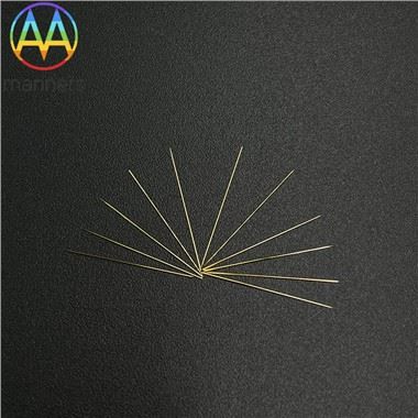
Echogenic Cannula Tips
Oct 01, 2026
Under live TRUS, one of the most maddening moments is watching the needle tip simply… disappear. In glandular tissue, behind scar, within periprostatic fat, a non-echogenic polished tip reflects almost nothing back to the probe. The operator is left estimating depth by hub markings and hope. Over-advance by a few millimeters and the apical seed lands in the urethra; angle slightly wrong and a posterior seed hugs the rectal wall. These are not dramatic errors - they are the slow, cumulative kind that show up later as elevated rectal dose on post-implant CT.
The physics is straightforward. An ultrasound image is built from acoustic impedance differences. A mirror-smooth steel tip reflects sound away from the probe; a ground-bevel with an echogenic treatment scatters returning echoes toward it, rendering the tip as a bright, trackable line. Add a hub bevel mark, and the operator also knows which way the tip is pointing before advancing - critical when placing peripheral seeds close to the capsule.
Echogenic tips are not monolithic. Flat-bevel tips offer a stable entry for template work. Long bevels give a more acute angle for shallow, fine approaches. Short bevels favor controlled deep puncture with less tissue spread. Echo-band tips add a marked ring a few millimeters back from the point for depth reference. Some premium needles combine an echogenic outer tip with a mirror-polished inner lumen, separating visibility outside from glide inside - two different surface goals on one instrument.
In practice, orientation discipline is everything. The bevel is turned toward the target margin so its echo face is presented to the probe. Scanning is done in both sagittal and axial planes; if the tip shadow hides a planned seed position, the probe is tilted 10–15° rather than guessing. No seed is deployed unless the tip echo and the planned coordinate coincide. For lung work under CT, the echogenic mark still helps during the brief US-confirmation step before final CT verification.
Experienced teams are candid: an echogenic tip does not make a poor scanner good. What it does is remove one variable so that skill can be expressed reliably. Centers that invested in probe technique and tip visibility report shorter learning curves for new implanters and fewer "rescan and reposition" loops. Conversely, teams that blame the image quality while ignoring bevel orientation rarely improve.
The philosophical point is simple: you cannot dose what you cannot see. Brachytherapy precision begins with tip awareness, not with the source activity. An invisible tip turns a geometric plan into a probabilistic guess.
Ahead, we can expect AI-assisted tip tracking that flags tip-echo loss in real time, augmented-reality overlays projecting the planned track onto the live scan, and automatic seed-shadow detection to warn of proximity to urethra or rectum. The echogenic tip is the analog foundation on which these digital aids will stand.