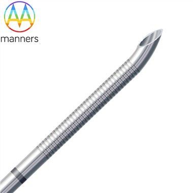
Core Sample Notch
Sep 24, 2026
Pain Points
In breast core needle biopsy (CNB), the sample notch is where diagnosis is either secured or lost. A notch that is too short yields fragmented tissue that cannot support hormone receptor testing or HER2 scoring. A notch with burred edges crushes ductal architecture, making it impossible for pathologists to distinguish invasive carcinoma from ductal carcinoma in situ (DCIS). A notch placed too far from the stylet tip makes ultrasound confirmation difficult, so the clinician fires before the trough is inside the lesion. Many low-cost breast biopsy needles copy the external shape of premium devices like Temno or Bard Magnum but ignore the critical relationship between notch volume, wall transition, and tissue settlement mechanics. The result is "adequate" samples that are actually insufficient for modern molecular subtyping.
Another widespread pain point is core loss during withdrawal. If the cannula inner wall is not properly electropolished and the stylet-cannula clearance is incorrect, the core smears along the lumen. Pathologists receive a rolled, hemorrhagic fragment instead of a cylindrical breast core. For 14G/16G mainstream breast CNB, this forces repeat puncture, increases hematoma risk, and damages patient trust. Thin-wall construction helps increase lumen size, but if wall consistency is poor, the notch region becomes the weakest point and kinks during spring firing. Additionally, many factories offer only one notch length (typically 15–17 mm) for all gauge sizes, ignoring the fact that an 11G vacuum-assisted device needs a longer trough than an 18G micro-biopsy. Using the same notch for every gauge is like using the same bucket for every well-it either overflows or comes up empty.
Working Principle
The core sample notch is a precision-machined side window in the stylet. Tissue advances into the open trough while the stylet is stationary. When the outer cannula fires, its sharp distal cutting edge shears the tissue at the mouth of the notch, capturing a defined cylinder. Notch length, depth, and edge geometry decide sample volume. A 14G notch commonly targets 15–20 mm core length; 16G and 18G reduce volume but improve cosmesis; 8G/11G/13G maximize architecture for vacuum-assisted and large-mass cases.
Thin-wall 304/316 tubing increases inner diameter without increasing outer diameter, so the same gauge harvests a larger core-a critical advantage in breast tissue where preserving architectural integrity directly affects treatment planning. Precision grinding gives the cannula cutting mouth a controlled bevel angle, usually 15–30°, with symmetrical facets and no heel burr. Echogenic etching on the stylet tip and cannula does not change the notch mechanics, but it changes clinical accuracy: the sonographer sees the trough under ultrasound, confirms lesion overlap, and fires only when the sample notch is correctly seated. This "see-notch, then fire" workflow reduces repeat passes from an average of 2.1 to 1.3 per lesion in experienced hands.
Equipment Classification
Notch formation uses CNC wire EDM, laser slotting, or ground-and-lapped windows. EDM gives precise notch volume with excellent edge quality; laser is faster but needs recast control; grinding is common for disposable stylet windows. Classification by gauge and application: 8G/11G/13G (large-bore, thin-wall, vacuum-assisted breast biopsy); 14G/15G/16G (standard CNB, best balance of yield and trauma); 18G/20G (micro-lesion, superficial nodule, thin breast tissue); custom (coaxial, curved, adjustable 9–25 mm notch per 2D/3D drawing). Inspection equipment includes projection microscopes for notch length verification, micro-borescopes for lumen patency, tensile testers for stylet-cannula bond strength, and ultrasound phantoms for echogenic marking visibility validation.
Practical Guide
Define notch length by lesion size: 10 mm for small sonographic targets under 8 mm, 20 mm for mass-type lesions above 15 mm. Keep stylet-cannula radial clearance at 0.01–0.02 mm to prevent tissue ingress while allowing smooth firing. Deburr all notch edges; never leave rolled metal that can crush tissue. Electropolish the cannula inner diameter to Ra ≤0.2 µm to preserve core integrity during withdrawal. Place echogenic bands proximal and distal to the notch, not on the notch mouth only, to avoid interfering with tissue entry. For OEM projects, supply 2D/3D drawings with notch volume, bevel angle, depth marking, and hub interface dimensions. Validate with breast-tissue simulant before clinical use. Require ISO13485 lot records with notch dimension Cpk ≥1.33.
Real-World Experience
A breast center compared 14G needles from two suppliers over 200 procedures. Supplier A had correct OD but rough notch edges from stamping; core fragmentation rate was 22%, and three cases required surgical excisional biopsy due to indeterminate pathology. Supplier B used thin-wall 316L, EDM notch with polished edges, electropolished lumen, and echogenic double-band marking; fragmentation dropped to 6%. Second-look ultrasound showed the notch seated in the lesion before firing in 94% of cases, reducing repeat passes. In vacuum-assisted 11G cases, thin-wall construction lifted usable core length by approximately 18% without increasing hematoma calls. A separate study of 16G CNB for microcalcifications found that EDM-notched stylets yielded 31% more intact architectural fragments than laser-notched equivalents, directly improving DCIS grading accuracy.
Summary and Elevation
The notch is not a hole in the needle; it is the specimen chamber of the pathology pipeline. Good breast biopsy design treats notch volume, edge quality, thin-wall inner diameter, and ultrasound marking as one integrated system. When these elements align, the pathologist receives architecture instead of debris; the surgeon gains confidence; the patient endures one pass instead of three. The best manufacturers treat the notch as a precision optical and mechanical component, not as a punched window.
Outlook and Recommendations
Adjustable notch devices will dominate small-lesion practice within five years. Notch volume will be matched to lesion subtype through AI-assisted ultrasound characterization: 10 mm for MICRO, 20 mm for mass, custom troughs for radial scars and papillary lesions. OEMs should standardize echogenic notch markers and validate "see-notch, then fire" workflows in training programs. Future breast CNB will be image-led, specimen-aware, and gauge-personalized, with notch geometry algorithmically generated from preoperative imaging.