Common Placement Errors And Clinical Risk Prevention Of Intraosseous Needles
Sep 28, 2026
1. Clinical Pain Points of Placement Errors
Non-standard intraosseous needle placement is a leading cause of preventable complications in emergency IO access, severely compromising rescue safety and treatment efficacy. Most frontline operators rely on empirical placement rather than standardized landmark positioning, resulting in frequent operational errors such as oblique penetration, excessive depth and incomplete cortical puncture. Incomplete bone cortex penetration leads to needle loosening and fluid extravasation, causing subcutaneous swelling, soft tissue injury and even compartment syndrome in severe cases. Excessive vertical penetration will pierce the posterior bone cortex, damaging deep soft tissues, blood vessels and nerves, and triggering persistent local pain and hematoma. For pediatric patients with fragile growth plates, inaccurate placement may cause epiphyseal injury and affect long-term bone development. In addition, repeated blind placement attempts enlarge bone puncture holes, increase the risk of postoperative osteomyelitis and cellulitis, and waste precious emergency rescue time.
2. Standard Placement Working Principle
Scientific intraosseous needle placement follows the core principle of landmark positioning, vertical penetration and resistance judgment, matching the physiological structure of human bone marrow circulation. The IO space maintains rapid and stable blood perfusion even under shock and low-pressure conditions, serving as a reliable central circulation channel. Standard placement requires accurate selection of safe bone landmarks, vertical alignment of the needle body with the long axis of bone, and uniform rotary penetration relying on the screw tube structure of medical intraosseous needles. Operators judge successful cavity entry through the obvious resistance loss sense after penetrating the cortical bone. Reasonable placement depth ensures the needle tip is stably located in the medullary cavity, avoiding shallow loosening and excessive penetration. Standardized placement maximizes the advantages of rapid drug and fluid delivery of IO access, ensuring infusion efficiency consistent with intravenous access while eliminating placement-related safety risks.
3. Placement-Adapted Equipment Classification
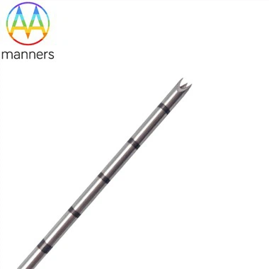
According to clinical placement difficulty and scenario adaptation, intraosseous needles are divided into three targeted placement types. Standard 15G universal placement needles adopt integrated screw tube and handle design, with moderate rigidity and penetration force, suitable for conventional landmark placement in adult and older child emergency scenarios, featuring stable vertical penetration and low placement error rate. Pediatric placement-specific fine needles are optimized in needle body length and tip radian, adapting to shallow cortical bone and fragile growth plate structure of children, effectively avoiding over-penetration and epiphyseal injury during placement. Custom placement needles support personalized size and structural adjustment based on 2D/3D drawings and sample customization, adapting to special placement scenarios such as skeletal deformity, obesity and local bone hyperplasia, solving placement failure caused by mismatched standard equipment. All products are made of medical stainless steel and processed by laser cutting and electropolishing to ensure smooth and accurate placement operation.
4. Standard Needle Placement Operation Guide
Clinical intraosseous needle placement implements standardized seven-step operational specifications to eliminate error risks. First, preoperatively confirm contraindications, avoid placement at fracture sites, open wounds and previous IO puncture points, and select safe sites including proximal tibia and distal femur. Second, disinfect the local skin and keep a sterile surgical field. Third, select matched needle specifications according to patient age and bone thickness, ensuring at least 5mm of the needle body exposed outside the skin to guarantee effective medullary cavity entry. Fourth, fix the bone landmark stably, keep the needle perpendicular to the bone surface, and rotate the handle evenly for slow penetration. Fifth, stop operation immediately when feeling resistance disappearance to prevent posterior cortex penetration. Sixth, aspirate a small amount of bone marrow to verify accurate placement in the medullary cavity. Seventh, fix the needle body tightly to avoid postoperative displacement and loosening.
5. Practical Placement Clinical Experience
Long-term emergency clinical practice verifies that standardized placement specifications can reduce intraosseous needle operational error rate by 90% and eliminate severe placement complications such as compartment syndrome and osteomyelitis. Standard vertical placement and resistance judgment technology increase the one-time placement success rate from 72% of empirical operation to 99%. Pediatric special placement needles completely avoid growth plate injury in child rescue, with zero developmental adverse events in clinical application. Customized placement needles solve the placement difficulty of special anatomical patients, realizing accurate cavity entry in complex scenarios. Strict placement contraindication implementation avoids repeated puncture and cross-site placement damage, significantly improving the safety and stability of emergency IO infusion treatment.
6. Summary and Refinement
Standardized placement operation is the core guarantee for safe and effective application of intraosseous needles. Most clinical adverse events of IO access stem from non-standard empirical placement rather than equipment performance defects. Accurate landmark positioning, vertical penetration and scientific resistance judgment form a complete safe placement system, which can give full play to the rapid and stable advantages of medullary cavity infusion, avoid tissue damage and infectious complications. Matching differentiated placement equipment with scenario-based operational standards realizes the organic unity of emergency rescue efficiency and clinical safety.
7. Clinical Standardization Optimization Suggestions
Medical institutions should establish unified intraosseous needle placement operation specifications and contraindication guidelines, standardize site selection, penetration angle and depth judgment standards. Strengthen special training for emergency operators, eliminate empirical blind placement habits, and improve the proficiency of standardized placement. Equip differentiated placement needle specifications for different age groups and special patients to realize precise equipment matching. Industry associations should compile placement operation teaching manuals, promote standardized placement technology in grassroots medical institutions, and comprehensively reduce the incidence of placement-related clinical complications.