The use of arthroscopes for arthroscopy
Nov 08, 2022
The miniaturization of the arthroscope has simplified its operation to outpatient and local anesthesia, but we still use the common knee arthroscope, which is performed in the operating room. The procedure is outlined below.
Under continuous epidural anesthesia, the affected limb was placed on the operating table, the epidural puncture needle was used to perform joint puncture on the suprapatellar sac, the exudate was extracted, and normal saline was injected into the joint to expand the joint cavity (the suspension height of the salt bottle was generally about 1m higher than the knee joint). Puncture points were selected at the center of the triangle formed by the lateral edge of the patellar tendon, the anterior edge of the lateral femoral malleolus, and the superior edge of the tibia. First, a small incision of about 0.5cm was made in the skin, and then a trocar matching the diameter of the arthroscope was used for puncture. The sharp occlusive device was removed and replaced with a blunt occlusive device. The arthroscope was inserted into the joint cavity, and the observation sequence was as follows: Patellar plica patellofemoral joint - on the medial fossae (medial wall, mediopatellar plica, medial malleolus fossae surface) - the stock of the medial tibial articular (medial meniscus, femoral medial malleolus and relative tibial articular surface below the former) - again to the patellar sac - lateral tibial articular (lateral meniscus, femur before external ankle and relative bone close written below) - lateral crypt (the outside of the wall, and stocks Crypt surface of the lateral condyle of bone, muscle). The above can be photographed. Finally, a biopsy can be performed to wash the full fluid in the back, pull out the trocar, and close the skin incision. There are different views on whether hemostatic bands should be used in arthroscopy. The authors believe that it is better not to use tourniquet at the beginning of arthroscopy, so that the internal tissue structure of the joint can maintain a normal appearance and it is easy to identify normal or abnormal tissues. An hour or so of arthroscopic surgery (including membrane-clearing biopsy) is sufficient for diagnosis. If the therapeutic procedure is to continue, a tourniquet should be applied.
Synovial pathologic examination is an important step in the diagnosis of joint disease. Synovial biopsy is usually performed at the same time after arthroscopic examination. There are three methods for synovial biopsy: ① Blind examination: that is, after the arthroscopy is completed, the mirror is pulled out of the cannula, the biopsy forceps are inserted, and the head of the biopsy forceps is felt through the skin with the other hand, and the biopsy is performed. ② If special lesions have been seen under the microscope, the original lens can be withdrawn from the cannula and replaced with a small arthroscope with biopsy forceps, which can be performed under direct vision. ③ If the surgeon wants to keep the observed lesion in the field of vision, a biopsy can be performed through the second puncture orifice.
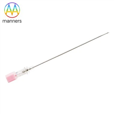
If you have any questions, please contact us. Our company can produce various customized needles, medical needles, puncture needles, hypodermic needles, biopsy needles, vaccine needles, injection needles, syringe needles, veterinary needles, pencil point needle, ovum pick up needles, spinal needles, etc. If you need customized needle products, please contact us. We look forward to your inquiry! The quality of the products manufactured in our factory will surely satisfy you!
Please contact us if you need: zhang@sz-manners.com