


Precision at the Tip: Ultrasound-Guided Nerve Blocks
Jul 23, 2024
Ultrasound examination is a diagnostic method that has emerged in the last 20-30 years and has gradually been applied to various clinical systems. However, ultrasound-guided nerve blocks have only appeared in recent years. In recent years, ultrasound technology has rapidly developed in functional evaluation and targeted therapy. It allows multi-directional scanning of target points from different angles, provides clear and easily recognizable images, and, compared to DR, CT, and MRI, it is non-radioactive, low-cost, and highly safe. More importantly, under ultrasound guidance, doctors can more precisely choose the puncture route, dynamically monitor the puncture path, instantly identify adjacent structures such as blood vessels, nerve plexuses, and pleura, guide the puncture needle to the treatment target, and observe the spread of local anesthetics. This significantly reduces the incidence of complications such as nerve plexus injury, vascular injury, and pneumothorax.
Ultrasound-guided peripheral nerve blocks can effectively relieve pain, and their procedures are simple, accurate, and require less medication. Currently, portable ultrasound technology enables timely and effective blocks of the brachial plexus and its branches, the lumbosacral plexus and its branches, the intercostal nerves, and more. It has become one of the key technologies in advancing medical techniques.
Applications
Ultrasound-guided peripheral nerve blocks are suitable for treating various pain locations.
Procedure Steps
- Based on the pain location, select the nerve that needs to be blocked.
- Perform the block under ultrasound guidance: Choose the appropriate ultrasound probe and the best ultrasound imaging mode to clearly display the peripheral nerve to be blocked. Determine the appropriate puncture point and path. Sterilize the local skin as usual, wrap the ultrasound probe with a sterile cover or sterile glove, and guide the puncture needle tip to the perineurium under ultrasound guidance. Inject an appropriate amount of local anesthetic (lidocaine or ropivacaine) to surround the nerve, then withdraw the needle to complete the procedure. During nerve block operations, ensure that the needle tip's position does not inject the drug into the nerve bundle, avoiding secondary nerve injury.
Indications and Localization Methods for Ultrasound-Guided Nerve Blocks
(1) Interscalene Brachial Plexus Block
Indications: Pain in the upper arm and shoulder.
Method: Place the patient in a supine position with the head turned to the opposite side. Position a high-frequency probe on the neck to display the interscalene groove, showing the brachial plexus ultrasound image (Figure 1).

(2) Axillary Brachial Plexus Block
Indications: Pain in the distal upper limb (hand and wrist).
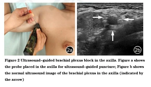
Method: Place the patient in a supine position with the axilla fully exposed. Position a high-frequency probe in the axilla to display the axillary artery and axillary vein. The "web-like" structure beside them is the brachial plexus (Figure 2), which includes the terminal nerves: median nerve, ulnar nerve, radial nerve, and musculocutaneous nerve.
(3) Suprascapular Nerve Block
Indications: Shoulder pain.
Method: Place the patient in a sitting or prone position. Position a low-frequency probe transversely on the middle and outer third of the scapular spine to display the scapular spine and suprascapular artery. The suprascapular nerve is located beside it (Figure 3).

(4) Intercostal Nerve Block
Indications: Intercostal neuralgia.
Method: Place the patient in a supine or lateral position. Position a high-frequency probe vertically on the ribs at the posterior or mid-axillary line to display the intercostal artery and vein. The low-echo structure beside them is the intercostal nerve (Figure 4).

(5) Femoral Nerve Block
Indications: Pain in the anterior thigh, knee joint, and inner side of the lower leg and foot.
Method: Place the patient in a supine position with the inguinal region fully exposed. Position a high-frequency probe at the midpoint of the inguinal region to display the femoral artery and femoral vein. The "web-like" structure beside them is the femoral nerve (Figure 5).

(6) Sciatic Nerve Block
Indications: Pain in the posterior thigh, posterior and lateral lower leg, and foot.
Method: Place the patient in a prone position, with the abdomen elevated if necessary. Position a low-frequency probe on the inner side of the greater trochanter to display the piriformis muscle. The "web-like" structure in the deep layer is the sciatic nerve. Alternatively, position a high-frequency probe above the popliteal fossa to display the popliteal artery and vein. The "web-like" structure in the superficial layer is the sciatic nerve, which can be traced upwards to the piriformis muscle (Figure 6).

(7) Common Peroneal Nerve Block
Indications: Pain in the lateral lower leg.
Method: Place the patient in a lateral position with the knee slightly bent and the affected limb on top. Position a high-frequency probe horizontally below the fibular head to display the elliptical structure, which is the common peroneal nerve (Figure 7). If the common peroneal nerve cannot be identified, position a high-frequency probe transversely above the popliteal fossa to display the sciatic nerve. Trace the sciatic nerve downward, where it divides into two branches. The lateral branch is the common peroneal nerve, which can be followed along its short axis to the fibular head for easier identification.

(8) Tibial Nerve Block
Indications: Pain in the posterior lower leg.
Method: There are two common blocking methods:
- Position a high-frequency probe transversely above the popliteal fossa to display the sciatic nerve. Trace the sciatic nerve downward, where it divides into two branches. The medial branch is the tibial nerve. Use real-time ultrasound guidance for needle insertion and injection.
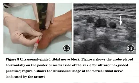
- Position a high-frequency probe transversely on the posterior medial side of the ankle to display the posterior tibial artery and vein. The "web-like" structure beside them is the tibial nerve (Figure 8).
Ultrasound-guided nerve block is a convenient, real-time, dynamic, multi-directional, and radiation-free diagnostic and therapeutic method. It has been proven to have high sensitivity and specificity as a visualization technique. This technology uses ultrasound imaging to clearly differentiate tissue structures and guide the puncture needle precisely to the target, changing the traditional blind puncture and probing method. It makes the operation more accurate, increases the success rate of puncture, reduces the number of punctures and operation time, and lowers the incidence of related complications such as nerve injury, inadvertent vascular entry, and bleeding, thereby enhancing medical safety.


