Lung mass biopsy needle method
Dec 13, 2021
First, the puncture area was routinely disinfected, then sterile towels were spread, and local infiltration anesthesia was carried out. (Be careful not to stab lung tissue during anesthesia). Lung biopsy can be divided into fine needle biopsy cytology and cutting needle biopsy histology.
1. The fine needle puncture cytological examination Needles Pierce the skin along the preoperative imaging first set by the puncture, direction and depth, slowly advance the needle (under ultrasound guided puncture the ideal), until to puncture, pull needle core, 20 ml syringe is connected, make needlepoint in keeping the negative pressure condition on the lesions within a small range, the 2 ~ 3 times, Then remove the negative pressure and pull the needle. Immediately push the aspirate onto the slide, and fix it with 95% alcohol after smear and send it to cell chamber for examination.
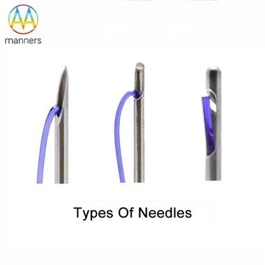
2. Histological examination of perforated reverse crochet needle puncture: first, make a small incision on the skin of the puncture point with a scalpel to facilitate needle insertion. After inserting the needle directly into the bottom of the tumor from the intercostal area along the original puncture direction and depth, the needle core was pulled out and the needle was inserted into the tumor for several times to make more tissues fall into the needle hole (the needle should be inserted and removed vertically during operation without swinging), and then the needle was pulled out. Push tumor tissue onto sterilized filter paper with needle core, fix and send for examination.
3. Trough puncture histological examination When the trough puncture needle with needle core is used for biopsy, it is different from the porous reverse crochet needle in that when the puncture enters the tumor capsule, the needle core is pulled out and the tumor tissue can be embedded into the needle cavity along the needle groove. At this time, the tumor tissue should be cut by rotating 180° ~ 360°, and the needle can be removed quickly. Push tissue block onto sterilized filter paper with needle core, fix and send for inspection.
Please contact us if you need: zhang@sz-manners.com