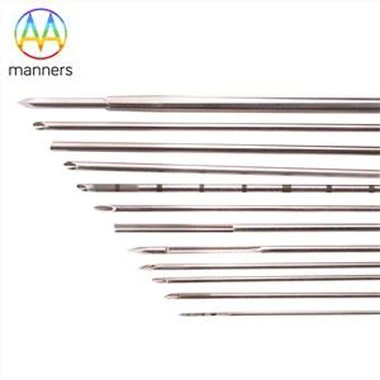
Epidural puncture needle block puncture method
Apr 02, 2022
1. Blind puncture method Epidural puncture can be done by straight and lateral method. The spinous processes of the cervical, upper thoracic and lumbar vertebrae are parallel to each other, and the straight-in approach is often advocated. For elderly people with calcified supraspinous ligaments and restricted spinal curvature, the lateral entry method is generally suitable.
Direct penetration method: A skin mound is made at the selected interspinous process space near the upper edge of the inferior spinous process, and then infiltrated layer by layer. The skin and ligament can be punctured with a 15 G sharp needle, and then the epidural needle is inserted along the needle hole. The needle location must be on the midsagittal line of the spine. The needle tip penetrates the skin, subcutaneous, supraspinous ligament, interspinous ligament, ligamentum flavum and other tissue layers. The resistance of penetrating the ligamentum flavum suddenly disappears, indicating that it has entered the epidural space.
Side entry method The side entry method is to insert the needle 1.5 cm next to the spinous process space, and puncture the needle at a 75° angle with the skin to pierce the interspinous process hole, avoiding the supraspinous ligament and the interspinous ligament, and enter through the ligamentum flavum. epidural space
Cannulation method for continuous epidural block: When it is determined that the epidural needle has entered the epidural space, the bevel of the needle is directed to the cranial or caudal side, and a graduated needle is inserted through the epidural needle lumen. 20 G catheter. The catheter should extend beyond the tip of the needle and into the epidural space by 3 to 4 cm. Secure the catheter with a sterile dressing. The catheter is placed to infuse the drug multiple times or continuously.
Drugs and dosage: First, inject a test amount of local anesthetic, generally 3-5 ml of 1.6%-2% lidocaine. Before injection, it must be sucked back to confirm that there is no blood and cerebrospinal fluid.
2. Puncture under ultrasound positioning Ultrasound positioning and guidance technology has been widely used in minimally invasive surgical anesthesia, regional block and vascular puncture, etc. It also provides a new method for epidural puncture and catheterization. High-quality ultrasound imaging can help the operator to quickly and accurately locate the epidural space, guide the puncture needle and catheter into the epidural space, and complete the epidural positioning tube, which can reduce the number of try-ons and reduce the discomfort of the patient. Improve patient satisfaction.
The operation steps are to first scan and mark the highest point of the iliac crest, then move the probe to the head to scan each lumbar intervertebral space and spinous process, and locate the third spinous process at the measured intersection. The epidural related structures were identified and the clearest gap was shown, and the puncture point was located approximately 1.0 cm from the midline in this gap. The puncture site was routinely disinfected, and after local anesthesia with 1% lidocaine, the puncture point was taken as the center of the probe, and a paramedian longitudinal section scan was performed.
Ultrasound sonography (from superficial to deep) shows that the most superficial layer is the skin, the fascia is linear hyperechoic, the adjacent spinous processes are “city wall-like”, the bone surface is arc-shaped linear hyperechoic, and the back is covered by acoustic shadows. At the top of the two spinous processes, due to the different orientations of the ligament fibers in each group, the ultrasound beam produces anisotropy, and the supraspinous ligament, interspinous ligament and interarch ligament are moderately low and uneven echo images. The dura mater and the ligamentum flavum have dense tissue structures, and the sonogram shows moderately hyperechoic bands, mainly the ligamentum flavum. Between the dura and the ligamentum flavum is the epidural space, and moderate to low echoes can be seen in the cavity. Epidural fat sonography (Figure 6). The operator inserts the epidural needle through the ligamentum flavum into the epidural space according to the acquired images of the intervertebral space. When the puncture needle reaches the ligamentum flavum, the resistance increases, withdraw the puncture needle core, connect the low-resistance syringe, and slowly inject the normal saline containing air bubbles; when the puncture needle breaks through the ligamentum flavum, no cerebrospinal fluid and blood are withdrawn, and the air bubbles are not compressed. The normal saline bolus injection without resistance confirms the successful puncture.
3. Puncture under C-arm positioning Under the C-arm fluoroscopy, the spinous process of the vertebral body can be clearly identified and the real-time position of the puncture needle can be clearly seen, which can improve the success rate of puncture.
If you have any questions, please contact us. Our company can produce various customized needles, medical needles, puncture needles, hypodermic needles, biopsy needles, vaccine needles, injection needles, syringe needles, veterinary needles, pencil point needle, ovum pick up needles, spinal needles, etc. If you need customized needle products, please contact us. We look forward to your inquiry! The quality of the products manufactured in our factory will surely satisfy you!
Please contact us if you need: zhang@sz-manners.com