Endoscopic Ultrasound (EUS)
Jul 30, 2024
Ⅰ Introduction
Endoscopic Ultrasound (EUS) is a fascinating technique that combines endoscopy and ultrasound technology. Since its introduction in the 1980s, EUS has undergone continuous improvements and has become an essential tool for diagnosing gastrointestinal (GI) diseases. It features an ultrasound probe at the tip of the endoscope, which allows it to capture real-time images of the GI tract and surrounding organs. EUS is particularly useful in detecting small lesions and diagnosing conditions in the upper GI tract and biliary system.

Ⅱ Basics
Definition
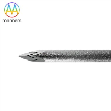
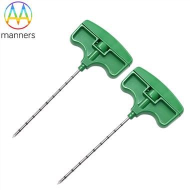
EUS is an integration of endoscopic and ultrasound imaging technologies. The ultrasound probe is attached to the tip of the endoscope, and depending on the relationship between the scanning plane and the endoscope, EUS can be classified into radial and linear types. Radial EUS has a scanning plane perpendicular to the endoscope's long axis, while linear EUS has a scanning plane parallel to it. Both types are used for disease diagnosis, but linear EUS also allows for fine-needle aspiration and treatments such as celiac plexus block and pancreatic cyst drainage. EUS is mainly applied in abdominal surgery, especially for diagnosing diseases of the upper GI tract, including the stomach and duodenum, as well as biliary and pancreatic diseases.
Principle
EUS combines real-time ultrasound scanning with endoscopic technology. The ultrasound probe at the end of the endoscope can perform radial or linear scanning. Radial scanning provides a broad view, making it easier to locate lesions, while linear scanning offers a more focused view, suitable for examining smaller organs. EUS is particularly useful in abdominal surgery for diagnosing diseases of the upper GI tract and biliary system. It provides detailed images of the GI tract layers and surrounding organs, significantly enhancing the detection of small lesions. Additionally, EUS-guided fine-needle aspiration can obtain tissue samples from areas such as the pancreas and liver.
Ⅲ Clinical Applications
Clinical Value
EUS is a commonly used medical examination method, particularly for detecting GI tract lesions, evaluating wall abnormalities, staging esophageal and gastric cancers, diagnosing varices, assessing malignant lymphomas, and diagnosing diseases of the pancreas, bile duct, and liver. However, EUS is not suitable for all patients. Those with severe mental disorders, significant cardiopulmonary diseases, suspected upper GI perforation, corrosive esophagitis, acute severe throat conditions, suspected thoracic aortic aneurysm, or severe spinal deformities are not ideal candidates for this procedure.
Examination Process
1. Preparation
Patients should fast for 12 hours before the examination. For upper GI EUS, the throat is numbed with a lidocaine spray, and diazepam may be administered. For colon EUS, laxatives are used to clear the bowel, and a cleansing enema is performed two hours before the examination.
2. Method
EUS can be performed using direct contact, water-filled balloon, or water immersion methods. If further examination is needed, the stomach can be rinsed and filled with de-aerated water for better ultrasound imaging.
3. Procedure
An electronic endoscope is used first for a thorough examination, followed by EUS for detailed inspection. The water-filled balloon method is used for the esophagus and surrounding organs, while the water immersion method is best for the stomach and nearby structures. For pancreatic examination, scanning starts from the tail and proceeds to the body and head of the pancreas.
Precautions
EUS may cause complications such as aspiration, bleeding, perforation, throat injuries, aspiration pneumonia, anesthesia-related incidents, and equipment-induced damage. EUS-guided fine-needle aspiration carries additional risks like pancreatitis, infection, and bleeding. However, the risk of needle tract seeding is lower compared to percutaneous methods. Pre-examination preparations should include coagulation tests, and defoaming agents may be used to improve image quality.
Ⅳ Report Interpretation
Clinical Interpretation
1. GI EUS Diagnosis
The GI wall appears in five layers under EUS, each corresponding to different structures and potential pathologies. For instance, polyps in the superficial mucosa, leiomyomas in the deep mucosa, and lipomas in the submucosa can be identified. Tumors like gastric cancer and lymphomas typically present as hypoechoic changes, affecting multiple layers. Ectopic pancreas in the gastric antrum is another common finding.
2. Pancreatic and Biliary Diseases
Proximal bile duct and gallbladder diseases can often be diagnosed with surface ultrasound, but distal bile duct, ampullary, and pancreatic head lesions are better visualized with EUS. EUS provides clear images of these areas, free from gas interference. Acute and chronic pancreatitis, pancreatic tumors, and bile duct stones are common conditions evaluated with EUS.
3. Tumor Staging
EUS is crucial for diagnosing and staging tumors, determining their depth of invasion, lymph node involvement, and surrounding organ invasion. It plays a significant role in preoperative staging, assessing resectability, evaluating treatment response, and detecting postoperative recurrence.
4. EUS Clinical Diagnosis
EUS provides precise anatomical localization, aiding in the differential diagnosis of raised lesions, tumor staging, biliary and pancreatic disease diagnosis, and EUS-guided interventional treatments.