Ultrasound-Guided Thermal Surgery
Oct 02, 2026
Ultrasound-Guided Thermal Surgery
There is a particular kind of silence in a well-run ablation room - not the sterility of an operating theater, but a focused quiet in which the only voices are the sonographer calling out depths and the faint click of the generator's foot pedal. It is in this silence that one realizes the uncomfortable truth veteran interventional gynecologists keep repeating: the hardest part of radiofrequency fibroid therapy is not generating heat, it is seeingwhere the heat goes. For all the marketing around "smart generators" and "precision electrodes," the limiting factor has never been wattage. It has been the human eye interpreting a flickering ultrasound frame while a needle tip hovers millimeters from a structure that must not be burned. Clinics discover this gap the hard way. A fibroid that appears as a tidy hypoechoic oval on a pre-op still image will, in live scanning, reveal a bulge toward the bladder dome, or a posterior lip that nearly kisses a mobile loop of rectosigmoid. Operators who trust the printed image over the moving one have learned costly lessons: a serosal thermal injury here, a transient thermal irritation of the bowel there, a patient who feels a sharp pang because the tip drifted half a centimeter closer than the plan allowed. The paradox of modern fibroid ablation is precisely this - the instrument is mature, the evidence is solid, yet confidence between rooms varies as widely as the skill of the hand on the probe.
To appreciate why imaging is the true co-surgeon, one must slow down on what the sound actually shows. B-mode ultrasound renders the metallic needle as a bright linear echo, sometimes with faint comet-tail artifact that, in experienced hands, becomes a depth ruler rather than a nuisance. As radiofrequency current drives ionic oscillation in the fibroid, tissue water heats, microbubbles form, and the nodule brightens into a hyperechoic cloud whose leading edge is, in effect, a thermal footprint. The operator is not guessing temperature; they are watching its shadow. Sustain the core above 60°C and smooth-muscle cells lose membrane integrity; hold the peripheral myometrium below the lethal threshold through measured ramping, and the womb keeps its viable shell. Later, contrast-enhanced ultrasound - microbubble contrast injected intravenously - maps the non-perfused zone, the "dead core" that no longer lights up. This visual feedback loop is what separates RF ablation from the older blind thermal era, when intracavitary heating or interstitial probes without imaging could only hope the energy had landed where intended. The needle delivers; the screen confirms; the margin is negotiated in real time.
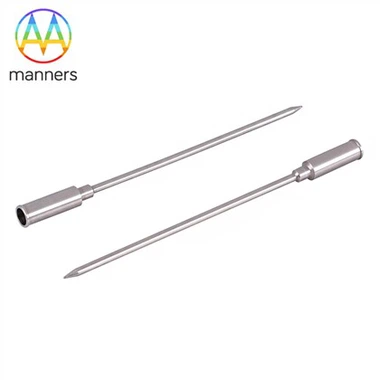
Devices engineered for image-guided work carry a set of quiet design decisions that non-specialists rarely notice until they are missing. The shaft must produce minimal acoustic shadow at the tip, or the very needle you are guiding becomes a black bar obscuring the target. Hub orientation matters - a bevel that faces the wrong way can make the tip appear to "jump" on screen. Laser-etched depth rings, once considered a luxury, have become near-essential: they let the operator translate the on-screen bright line into "tip at 2.0 cm, 2.5 cm, 3.0 cm" without mental arithmetic under time pressure. Insulation is not merely about patient safety; a poorly insulated shaft burns the track on entry, throwing a false hyperechoic streak that mimics a bubble cloud and misleads the scan. Generators, too, are part of the imaging choreography: monopolar systems require a dispersive pad placed so it does not crowd the probe angle; bipolar systems simplify the field for transvaginal routes; impedance roll-off and temperature cutoffs let the sonographer's eye stay on anatomy rather than darting to a wattage display. In truth, the needle and the ultrasound screen are not two instruments used in sequence - they are one instrument held in two forms.
The workflow in a disciplined room has almost the cadence of a ritual, though nothing mystical about it. First, the team decides between freehand needle guidance and a fixed-angle guide bracket - freehand offers flexibility for curved approaches, the bracket offers reproducibility for training. Then the viewing plane: long-axis to follow the entire shaft like a pencil line through the myometrium, or short-axis cross-sections to confirm depth at discrete points. Before the skin is punctured, someone verbalizes the safety margin: distance to serosa, to endometrium, to bladder, to any visible bowel loop. The bladder is deliberately managed - partially filled to create an acoustic window for a posterior lesion, or emptied to drop the dome away from an anterior one. Power is never "set and forget"; it is nudged in small increments as the hyperechoic cloud grows, paused if the rim approaches the serosal line, resumed after a scan. Anterior fibroids usually yield to a transabdominal track angled off the pubic symphysis; posterior and cervical lesions often favor a transvaginal approach where the needle travels a shorter, more controllable path; very large masses may require switching windows mid-procedure, the sonographer re-centering the probe while the operator holds steady. Every session ends not with a suture but with a triptych of images: pre-ablation baseline, intra-procedural bubble cloud, and post-ablation perfusion check - stored not for audit theater, but because an operator's learning curve is literally written in side-by-side frames.
Experience in this field accrues in millimeters and in warnings passed informally between mentors. Senior operators speak a dialect of distances: "tip 1.2 cm off the serosa, no closer," "thread through the avascular myometrial stripe," "core at 90°C, margin held under 42°C." They ablate a sector, freeze the frame, scan for any residual color Doppler blush, and add one more short track only if a viable rim persists. One documented giant-fibroid case - a lesion approaching 11 cm - used a multipoint moving ablation with immediate contrast assessment after each fan, refusing the temptation to declare victory on a single pass. The folklore that travels with them is consistent: the best ablations look boring. No sudden cavitation pop, no patient wince, no last-second repositioning. Drama at the table, they say, is almost always a sign that someone rushed the ramp or ignored a wandering bowel loop. Newer operators are also taught the patient-language part: to explain, before the first watt, that the ultrasound may still show a "shadow" for months after the tissue is biologically dead, so that a follow-up scan does not read as failure.
Lifting the gaze, the philosophical shift here is quietly profound. The radiofrequency needle does not replace the clinician; it amplifies the clinician's sight. The machine senses heat, but only the trained eye reads consequence - the millimeter of serosal margin, the drift of a bowel loop, the faint residual blush that means one more track. In an era infatuated with "smart" devices that promise to remove human error, image-guided thermal surgery insists on a humbler truth: technology extends judgment, it does not substitute for it. The hand on the probe remains the final arbiter.
Looking to the next decade, the recommendation is almost contrarian: invest first in ultrasound simulation and credentialing, not in yet another generator with a brighter screen. Build phantom trainers that reproduce the comet-tail artifact, the moving bowel loop, the posterior fibroid hugging the serosa, so juniors can fail safely before they touch a patient. Next-generation systems should fuse real-time B-mode with AI-assisted margin detection and generator throttling, warning the operator a half-second before the thermal front nears a forbidden structure. Yet even then, the discipline of the millimeter will belong to the human. Procurement should ask not "how many watts?" but "how well does this system teach my sonographer to see?" Because in thermal fibroid therapy, the most expensive component in the room will never be the needle - it will be the trained eye that guides it.