The Significance Of The Angle: How Curved Meniscal Repair Needles Reshape The Surgical Approach To The Posterior Horn
Apr 14, 2026
The Significance of the Angle: How Curved Meniscal Repair Needles Reshape the Surgical Approach to the Posterior Horn
Q&A Approach
When a meniscal tear in the posterior horn is hidden from direct view during knee arthroscopy, how can a surgeon precisely deliver repair instruments to this "blind spot"? Traditional straight needles, guided from the front, struggle to reach this "anatomical dead zone." The curved meniscal repair needle was born to solve this geometric dilemma. But how is the curvature calculated to balance optimal biomechanical repair with the avoidance of neurovascular structures?
Historical Evolution
The exploration of meniscal repair paths has evolved from "open visualization" to "arthroscopic direct access" and now to "curvilinear reach." In the 1980s, arthroscopic meniscal repair emerged but was largely limited to the middle and anterior portions. In 1995, the first 12-degree curved needle was used for posterior horn tears, raising success rates to 40%. By 2005, 24-degree curved needles combined with the "over-the-top" technique expanded the repairable range of the posterior horn by 60%. The advent of adjustable curved needle systems in 2015 allowed for real-time angle adjustments based on tear location. Today, personalized pre-bent customization based on patient 3D CT scans is turning the concept of "one key for one lock" into a reality.
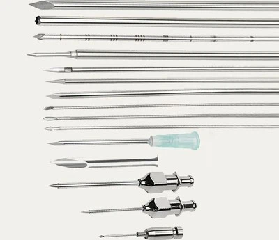
Curvature Geometry
Precise matching of needle tip angles to meniscal anatomy:
|
Curvature Angle |
Anatomical Region |
Puncture Path Characteristics |
Critical Structures Avoided |
|---|---|---|---|
|
0° (Straight) |
Body, Anterior Horn |
Shortest path; intuitive operation |
Patellar fat pad, Articular cartilage surface |
|
12° |
Lateral posterior horn, Posterior body |
Navigates anterior to femoral condyle rim; moderate lateral deflection |
Popliteus tendon (~8–10 mm from lateral meniscus posterior horn) |
|
24° |
Medial posterior horn apex, Root attachment |
Significant circumvention to reach hidden areas |
Popliteal artery (~5–7 mm from posterior capsule), Tibial nerve |
|
Reverse Curve |
Menisco-capsular junction |
Suture placement from outside-in |
Prevents suture from cutting through the meniscal body |
Optimization of Force Transmission
Biomechanical design of curved needle shafts:
Stress Distribution: A continuous, gradual curve (Radius ≥15 mm) is superior to an angular转折 (sharp corner), dispersing puncture stress evenly to prevent shaft fracture at the bend.
Torque Transfer: Nitinol's superelasticity ensures rotational torque from the handle is efficiently transmitted to the tip without angular loss.
Puncture Resistance: The penetration resistance of a 24-degree curved needle is ~35% higher than a straight needle, necessitating sharper tip geometry and surface coatings.
Suture Friction: Electropolishing (Ra ≤0.25 μm) of the curved lumen reduces suture friction by 60%.
Navigation and Visualization
Spatial localization techniques under arthroscopy:
30° Oblique Lens: Rotating the camera view combined with the curved needle creates a "mirror-image" operation, expanding coverage of the blind spot.
70° Wide-Angle Lens: Directly visualizes the posterior recess, providing a direct view for curved needle manipulation.
Probe Localization: Using a straight probe to identify the tear location first, then selecting a curved needle of proportional curvature to simulate the path.
Laser Markings: Laser-etched depth markings on the needle tip allow real-time assessment of penetration depth under the scope.
Clinical Repair Matrix
Needle selection strategies for different tear patterns (O'Connor Classification):
|
Tear Type |
Preferred Needle |
Assistive Technique |
Repair Success Rate |
|---|---|---|---|
|
Longitudinal (Red Zone) |
12° Curved |
Vertical mattress suture |
85–90% |
|
Radial Tear |
0° or 12° Curved |
Horizontal mattress + vertical reinforcement |
70–80% |
|
Bucket Handle |
Primarily 0° Straight, 12° Curved assist |
Inside-out all-inside technique |
90–95% |
|
Horizontal Cleavage |
24° Curved |
Separate sutures for superior/inferior leaves |
60–70% |
|
Root Tear |
24° Reverse Curve |
Transtibial pull-out fixation |
80–85% |
Safety Margin Control
Risk management for curved needle manipulation:
Vascular Distance: When using a 24-degree curved needle via the posteromedial portal, the mean distance from the needle tip to the popliteal artery must be ≥5 mm.
Nerve Protection: During lateral approaches, maintain a ≥8 mm safety distance between the needle and the common peroneal nerve.
Cartilage Avoidance: The arc trajectory of the needle tip should run parallel to the femoral condyle articular surface to avoid scratching the cartilage.
Real-time Monitoring: Use of force-sensing curved needles where a sudden spike in resistance signals potential contact with critical structures.
Chinese Anatomical Data
Measurements based on MRI of 500 Chinese adult knees:
Posterior Horn Depth: The medial meniscus posterior horn averages 9.2±1.8 mm from the posterior capsule.
Curved Path: Ideal penetration depth for a 24-degree curved needle is 25–35 mm.
Individual Variation: The posterior capsular space in female patients is on average 2–3 mm narrower than in males.
Obesity Impact: Joint space narrowing in BMI >30 patients may necessitate selecting needles with a smaller curvature.
Frontiers of Technological Integration
The intelligent future of curved needles:
EM Navigation: Needle tip integrated with sensors for real-time 3D position display.
Adjustable Curvature: Shape-memory alloys allowing remote-controlled angle adjustment during surgery.
Robotic Assistance: Robotic arms providing stable control of curved needles, eliminating hand tremors.
Augmented Reality: AR glasses overlaying neurovascular pathways for visualized safe corridors.
Pre-op Simulation: Finite element analysis based on patient MRI to simulate the optimal puncture path.
Economic Considerations
Value proposition of curved repair needles:
Technology Premium: 24-degree curved needles cost 40–60% more than straight needles.
Clinical Value: Enables arthroscopic repair of posterior horn tears that previously required open surgery.
Learning Curve: Curved needle operation requires an additional 20–30 cases of training but significantly expands the treatable case spectrum.
Social Benefit: Minimally invasive repair preserves meniscal function, preventing or delaying osteoarthritis.
Dr. James Lubowitz, Past President of the International Society of Arthroscopy, Knee Surgery and Orthopaedic Sports Medicine (ISAKOS), commented: "The significance of the curved meniscal repair needle is that it allows the surgeon's hand to 'reach around' anatomical obstacles to touch injuries that were once unreachable. Every successful posterior horn repair is a perfect application of geometry in medicine." Within the millimeter-level confines of the joint space, precise angular calculations are redefining the boundaries of meniscal repair possibility.