The Role Variations Of Chiba Needles In Different Tissue And Organ Biopsies
Jun 09, 2026
Biopsy is the gold standard for obtaining pathological diagnoses, and the Kashima needle is the "arrow piercing through the clouds" for precise biopsy of deep organs. However, the tissue characteristics, anatomical environment, and risks of different organs in the human body are completely different, which determines that the application strategy of the Kashima needle needs to be "tailored to local conditions." This article will take the lungs, liver, pancreas, and soft tissues of bones as examples, to compare and analyze the technical key points, advantages, challenges, and role differences of the Kashima needle in biopsy of different tissues and organs, and demonstrate its "precise tool" adaptability.
I. Lung Biopsy: A Precise Dance Between the "Air Sac" and the "Blood Vessel"
The lungs are filled with air and are rich in blood vessels. The main risks of biopsy are pneumothorax and bleeding.
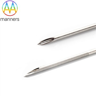
- Needle selection: The preferred choice is the 22G ultra-fine leaf needle. Its extremely thin diameter can minimize the tearing of the pleura and lung tissue to the greatest extent, significantly reducing the incidence of pneumothorax (especially when passing through the interlobar pleura). Even when passing through small blood vessels, the risk of bleeding is extremely low.
- Core technique: Emphasize "quick passage through the pleura, precise targeting of the lesion." During insertion, patients are required to hold their breath. The operator must be decisive and smooth in passing through the pleura, avoiding repeated adjustments of the needle tip within the lung (increasing pleural friction). For sub-centimeter nodules, the "coaxial technique" is often used: use the 22G leaf needle to precisely locate the nodule, introduce an outer cannula as a stable channel, and then use the biopsy gun through the cannula to take multiple samples, avoiding repeated puncturing of the pleura.
- Imaging guidance: CT is the gold standard, which can clearly show the relationship between the needle tip and the lesion, blood vessels, and interlobar fissures. For lesions close to the chest wall, ultrasound guidance is real-time and non-radiative, with obvious advantages.
II. Liver Biopsy: Safe Sampling in the "Blood Sinuses"
The liver has abundant blood supply and a soft texture. The main risks are bleeding and needle tract implantation.
- Needle selection: Either 20G or 22G needles can be used. The 20G needle can obtain a more complete tissue section, which is beneficial for pathological diagnosis (especially for small nodules in the context of liver cirrhosis), but there is a slightly increased risk of bleeding. The 22G needle is safer and is suitable for those with poor coagulation function or for lesions close to major blood vessels.
- Core technique: The key lies in precisely planning the puncture path, requiring it to pass through a certain thickness of normal liver tissue (to compress the needle track) and absolutely avoiding the hepatic major vessels and bile ducts. The puncture often uses the "rapid aspiration method": after the needle tip enters the lesion, the syringe is connected to maintain negative pressure, and several short and rapid punctures and retraction are made within the lesion to cut and obtain sufficient cells. Before withdrawing the needle, the negative pressure must be released to prevent the sample from being sucked into the syringe barrel.
- Image guidance: Ultrasound guidance is the preferred method due to its real-time nature and the ability to display blood flow (color Doppler), effectively avoiding blood vessels. CT is used for lesions that are unclear on ultrasound.
III. Pancreatic Biopsy: High-risk Procedures in the "Rich Blood Supply" and "Hidden" Categories
The pancreas is located deep within the body, surrounded by numerous large blood vessels, and has a high risk of pancreatitis. It is one of the most risky areas for biopsy.
- Needle selection: It is strongly recommended to use a 22G or thinner needle from Chiba. The core principle is to prioritize safety. Fine needles can significantly reduce the risks of bleeding, pancreatic fistula, and acute pancreatitis. The cytological diagnosis accuracy of fine needle aspiration is already very high in pancreatic cancer cases.
- Technical core: Pathway planning must be extremely cautious. Usually, transgastric or transintestinal puncture is chosen, using the gastrointestinal tract as a "buffer zone" to avoid direct damage to the peritoneum. The operator is required to be familiar with the three-dimensional anatomy of the surrounding pancreatic vessels. The technique must be extremely gentle to avoid rough operations. The "multiple points, small amount" sampling strategy is often adopted.
- Image guidance: Enhanced CT or endoscopic ultrasound (EUS) guidance is the mainstream. EUS-guided puncture paths are shorter, can avoid most blood vessels, and can perform biopsy on pancreatic head and body lesions in the posterior part of the stomach, which has become an important method.
IV. Bone and Soft Tissue Biopsy: Overcoming the Challenges of "Hardness" and "Mobility"
The bones are hard, the soft tissues have mobility, and there are different requirements for the rigidity and control of the needles.
- Bone biopsy: A bone puncture needle with sharp cutting edges or a specially designed one (considered as an "enhanced variant" of the Chiba needle) should be used, or a "drill-pull" combination can be employed. The Chiba needle is mainly used for aspirating soft tissue components in osteolytic lesions with abundant soft tissue. The operation needs to be precisely planned under CT guidance to avoid nerve and blood vessel damage.
- Soft tissue biopsy: For muscle or subcutaneous masses, ultrasound guidance is the preferred method. The needle tip movement and the deformation of the mass can be observed in real time. The mass needs to be "fixed" with the needle tip before sampling to prevent it from sliding. A 20G needle can be used to obtain tissue strips.
Conclusion
The application of Kiyama needles in various organ biopsies represents a "tailored" guideline for precision medicine. In the lungs, it is a "sleuth" striving for "extreme fineness" to pass through the air sacs; in the liver, it is an "explorer" ingeniously planning the path to avoid the blood sinuses; in the pancreas, it is a "craftsman" walking on thin ice, prioritizing safety above all else; in bones and soft tissues, it is a "multi-talented individual" capable of adapting its form to different hardnesses. A profound understanding of these differences is the fundamental prerequisite for clinicians to safely and effectively obtain high-quality pathological specimens, thereby formulating correct treatment plans for patients.