The Precision Engineering Behind The 14G Core Biopsy Needle
Jun 16, 2026
A seemingly ordinary 14G core biopsy needle is, in reality, a crystallization of materials science, precision machining, and bioengineering expertise. Its卓越 (exceptional) performance stems from the relentless pursuit of perfection in every manufacturing detail.
I. Material Selection: The Triangular Balance of Strength, Sharpness, and Biocompatibility
As outlined in your provided materials, stainless steel and titanium alloys are the two mainstream material choices.
- 304/316L Stainless Steel: This is the classic choice for medical-grade steel. It offers outstanding corrosion resistance, withstanding autoclave sterilization without rusting. Its strength and hardness support the manufacturing of thin-walled cannulas, allowing the 14G needle to maintain a 2.1 mm outer diameter while maximizing the inner lumen (approximately 1.6 mm ID) to accommodate a thicker tissue core. Its drawback is a higher specific gravity, resulting in a heavier feel.
- Titanium Alloy: Represented by Ti-6Al-4V, it boasts an exceptional strength-to-weight ratio. A 14G needle made of titanium alloy can be over 40% lighter than its stainless-steel counterpart. This offers a significant advantage during prolonged, high-precision manual procedures, notably reducing clinician hand fatigue. Additionally, titanium provides superior biocompatibility and is non-magnetic, making it suitable for MRI-guided biopsies. However, it is more difficult to machine, costlier, and slightly less wear-resistant than stainless steel.
- Medical Polymers: Primarily used for the hubs or handles of disposable, low-cost biopsy needles. For instance, Polycarbonate offers excellent transparency for visualizing blood return, while ABS plastic provides toughness and impact resistance. The needle shaft itself is rarely made of plastic, as it cannot provide sufficient puncture force.
II. Needle Tip Geometry: The First Gatekeeper of the Puncture Experience
The tip of a 14G core needle is not a simple bevel but a meticulously designed "cutting edge."
- Bevel Angle: Common tip bevel angles range between 15° and 30°. Smaller angles produce sharper tips with less resistance penetrating skin and fascia, but the tip is more prone to bending. The 14G needle typically employs an 18–22° compound bevel to balance sharpness with structural strength.
- Double/ Triple Bevels: To further reduce insertion resistance, premium tips feature secondary or tertiary bevels. This design allows the tip to "slice" through tissue like a scalpel rather than "tearing" it, minimizing tissue displacement and patient pain.
- Echogenic Markers: Near the tip, micro-pits or roughened surfaces are created via laser etching or sandblasting. These structures generate stronger echoes under ultrasound, allowing the clinician to clearly visualize the needle tip position.
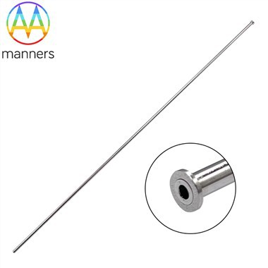
III. Precision Fit of the Inner Notch and Outer Cannula
This is the core mechanical assembly of the core needle, with tolerances controlled at the micron level.
- Inner Notch: Typically U-shaped or V-shaped, the surface undergoes mirror polishing. This smooth finish minimizes friction between the tissue and the notch wall, ensuring the tissue core can be extracted cleanly without adhesion or fragmentation.
- Outer Cannula: The leading edge of the outer cannula is also sharpened. The radial clearance between the inner notch and the outer cannula is merely a few microns. When fired at high speed, the outer cannula acts as a sharp blade, cleanly severing the tissue along the edge of the notch to form a perfect cylinder. Excessive clearance results in tissue crushing or "chewing"; insufficient clearance causes jamming.
- Spring Mechanism: The core driver of the high-speed ejection is a pre-compressed spring. High-quality springs, made from piano wire or oil-tempered spring steel, undergo rigorous fatigue testing to ensure stable firing speed and force after thousands of cycles. The firing force is typically maintained between 10–30 Newtons-sufficient to cut tissue without causing excessive needle vibration.
IV. Coatings and Surface Treatments
- Silicone Coating: Applying an ultra-thin layer of medical-grade silicone oil to the outer shaft significantly reduces friction during insertion, resulting in smoother advancement and less patient discomfort.
- Hydrophilic Coating: This becomes extremely slippery upon contact with water, outperforming silicone oil. It is often used for complex procedures requiring multiple adjustments of the needle angle.
- Anti-Reflective Coating: A black coating applied to the inner lumen reduces light reflection, facilitating visual inspection for blood return or tissue fragments under a microscope.
Conclusion
A small 14G core biopsy needle encapsulates knowledge from metallurgy, fluid mechanics, precision mechanics, and surface chemistry. It is these invisible aspects of precision engineering that ensure its reliable clinical performance. Every qualified 14G needle represents the perfect synergy between industrial manufacturing capabilities and medical demands.