The Clinical Logic Of IO Needles In Critical And Emergency Cases And Why Device Reliability Is Not An Option
May 28, 2026
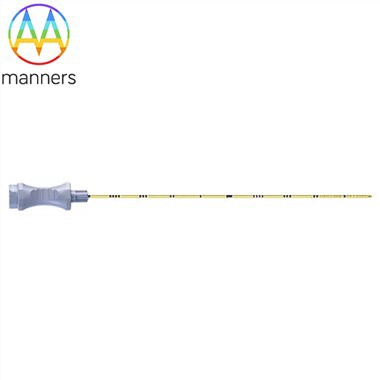
-- Intraosseous Access Needles + Manners: When veins disappear, the bone marrow becomes the last remaining blood vessels.
The American Heart Association (AHA) and the Pediatric Advanced Life Support (PALS) guidelines have an unwritten rule: if a venous access cannot be established within 90-120 seconds, an alternative route must be initiated - and in scenarios such as collapse shock, cardiac arrest, and massive bleeding from multiple injuries, peripheral veins are not "difficult to find," but rather do not exist at all (due to collapsed blood vessels, low blood pressure, skin edema, and severe burn contractures). At this point, the IO needle is not an "alternative," but the only feasible intravascular access available at that time.
1. Why Can the Bone Marrow Cavity Be Used as a "Blood Vessel"?
The sinusoidal veins within the bone marrow cavity are not directly equivalent to veins, but they are connected to the central circulation through a large, low-resistance network of cavities. After injecting drugs/liquids into the marrow cavity:
Efficacy time ≈ Central venous administration (usually a circulatory response should be observed within 30-60 seconds)
Can be infused with crystals, colloids, whole blood/blood products, epinephrine, amine vasopressors, sedatives, antibiotics - almost all IV drugs can be administered via IO as well.
Physiologically, the bone marrow cavity is a "non-collapseable rigid space" - unlike peripheral veins which can be compressed by shock, which is why in cases of severe hypovolemia, IO is actually more reliable than IV.
II. Five "Must Use IO" Battlefields
Scenario: Why IV fails? The value of IO
Out-of-hospital / In-hospital cardiac arrest: No pulse → Peripheral veins collapse; Difficult to maintain CPR position for long periods and unable to palpate blood vessels. Tibial proximal can be punctured within 10 seconds, and drugs can directly enter the central circulation.
Severe trauma / Hypovolemic shock (blood loss shock): Massive fluid resuscitation + collapse of blood pressure → Veins disappear; Outdoor environment with poor lighting. No need to palpate the pulse; Just touching the bone is sufficient - bones won't hide.
Pediatric emergencies: Infants and young children have extremely thin veins and blood volume is only ~70–80 mL/kg. IV attempts take as long as the blood loss time. PALS explicitly supports IO as the preferred rapid access route; Tibial proximal is the most stable.
Large-area burns / Severe edema / Obesity: Distorted body surface landmarks, veins obscured by edema or burned. IO should be "punctured downward to a hard area," bypassing the uncertainty of the body surface.
Disaster / Military rescue / During transport: Bumpy environment, fatigue of personnel, limited equipment. Manual IO (spring/rotational) can operate without power supply and is the most resistant to environmental interference.
Clinically, a common saying goes: "IO does not replace good IV, but when good IV is absent, IO can buy back some time for the patient's life."
III. The Common Requirement Behind the Three IO Driver Architectures: The Pins Must Be Robust.
Today, the clinical IO ecosystem is divided into three main paths. However, regardless of which path is chosen, the mechanical reliability of the needle itself is the ultimate limit:
A. Manual / Jamshidi-style (Manual / Jamshidi-type)
The surgeon holds the handle, inserts the needle perpendicularly into the bone surface, and rotates it forward - like tightening a wood screw. The advantages are quiet, wireless, and the cheapest; the fatal weakness is that for extremely dense adult bone, a lot of arm strength and hand feel training is required, and when fatigued, the torque becomes unstable → slipping → the needle bends / a groove is dug out on the bone surface → failure.
B. Battery-powered Drill Type (Powered / EZ-IO Style)
The reusable battery handle can hold the disposable needle group. The trigger activates the motor, which rotates and propels. The insertion time can be compressed to less than 10 seconds, with a high success rate (reported in the literature ~95%). However, the requirements for the needle body are as follows: The torque transmission must not cause the Hub to separate from the tube body, nor allow the core to retract - all interface connections must be resistant to torsion.
C. Spring-loaded (Spring-activated / BIG Type)
Pulling the trigger releases the stored spring energy, causing the needle to pierce through the cortex with a "bang." The advantage is that it is extremely fast and battery-free; the disadvantage is that the impact force is significant → the needle body must withstand transient axial loads without bending, flattening, or retracting the core.
The three are all converging towards the same goal: The IO pin withstands complex loads at critical moments - axial pressure + rotational torque + intermittent impact + bone debris friction - and it cannot bend, cannot retract the core, cannot get clogged, and cannot break.
IV. "Loss of Resistance" Feel: The Equipment Must Assist the User and Must Not Cause Any Disruption.
The success indicator for IO is not "seeing the blood flow back" (only 80–85% can be successfully extracted; failure to extract ≠ incorrect position), but rather:
Abrupt loss of resistance (pop / sudden change in tactile sensation): The tactile sensation that occurs at the moment of the cortex rupture.
The needle can stand upright (can remain upright on the bone surface without being pulled out).
No bulge in rapid saline infusion (flush test).
Among these three points, the rigidity of the needle, the clean cutting of the needle tip edge (without bone debris reaching the tube opening), and the zero loosening of the core-pipe fit determine whether the sensation is overwhelmed by "noise." A bent, blocked, or shaky IO needle will create a false drop (the bone surface is scraped with grooves, not a true penetration) or no drop (the needle tip is worn out, causing slippage on the cortical surface), directly leading to over-penetration or repeated attempts - and the patient is in the fourth minute of cardiac arrest.
V. Pediatrics: The Group Least Capable of Making Mistakes
The cortical bone at the proximal end of the baby's tibia may be only 1–2 mm thick, and the anterior-posterior diameter of the medullary cavity is also relatively shallow. The key points for the design of the IO needle in this area become:
Depth-limiting mechanism (adjustable retaining ring / stepped needle geometry): physically prevents over-penetration.
Fine specifications but continuous (gradual reduction in diameter at the 15G/18G transition section).
Sensation amplification: The needle must be cut cleanly - dull = more force applied = going beyond the thin dermis = into the joint/growth plate area.
This is why the IO needles are not simply "the same type of needle with a shorter tube": the pediatric specifications form an independent geometric system.
Conclusion
The clinical logic of the IO needle can be summarized in one sentence: Its existence is not for "needle puncture technique," but to eliminate the single point failure of "being unable to administer medication if a vein cannot be found." Within that 90-second window, the needle's rigidity, cutting efficiency of the blade, tightness of the core passage fit, and surface smoothness - these micron-level parameters that Manners locked in the workshop through neck reduction, die forging, grinding, and electrolytic polishing - are the dividing line between clinical success and repeated failure. The equipment can be selected as manual/electric drill/spring, but "the needle not bending, not blocking, not sliding, not loosening" is the common prerequisite for all options.