The Century-Long Evolution Of Meniscus Treatment Philosophy — From Total Resection To Suture Whenever Possible
Apr 15, 2026
The Century-Long Evolution of Meniscus Treatment Philosophy - From "Total Resection" to "Suture Whenever Possible"
The history of meniscus injury treatment is a chronicleof transformation - from crude resection to meticulous repair, from short-term symptom relief to long-term joint preservation. Spanning over a century, this evolution mirrors medicine's fundamental shift from shallow understanding to deep insight, and from blind intervention to precision treatment.
Phase One: Cognitive Void and the Era of Total Resection (1885–1950s) - Groping in the Dark
In 1885, British surgeon Thomas Annandale performed the first documented meniscus operation. Yet, for over half a century, the fateof the meniscus was brutally simple: once injured, it was almost invariably removed in its entirety.
Medical understanding at the time was fundamentally limited. The meniscus was regarded as an "evolutionary remnant" or "muscular vestige," analogous to the appendix, believedto have little function. More critically, surgical techniques lacked the capability to manage tears while preserving the tissue. Open surgery offered limited visualization, making precise suturing impossible, and total resection became the only viable option.
In 1936, American orthopedic surgeon Don King summarized the prevailing view in a leading journal: "The meniscus is a non-functional remnant; patients usually recover well after excision and can return to sports."This mindset guided an entire generationof orthopedic practice.
However, discordant notes began to emerge. Patients who underwent total meniscectomy started experiencing progressive knee pain, swelling, and dysfunction 5–10 years postoperatively. Radiographs revealed classic osteoarthritic changes: joint space narrowing, osteophyte formation, and subchondral sclerosis. Yet, the dominant interpretation was that these patients were "predisposed to arthritis," rather than attributing the outcome to the surgery itself.
Phase Two: Dawn of Partial Resection (1950s–1970s) - Rediscovering Functional Importance
The 1950s brought pivotal studies that changed the meniscus's fate. In 1954, Fairbank published a milestone paper systematically describing postoperative radiographic changes after meniscectomy, known as Fairbank's triad: flattening of the femoral condyles, joint space narrowing, and osteophyte formation. He explicitly linked these changes directly to the absence of the meniscus.
Concurrently, breakthroughs in biomechanics quantified the meniscus's role. In 1968, Walker et al. demonstrated that the meniscus transmits approximately 50% of the load at full extension, rising to 85% at 90° of flexion. Removing the meniscus increases articular cartilage pressure by 2–3 times.
These findings gave rise to a new philosophy: shifting from "total resection" to "partial resection." The idea was to remove only the torn segment while preserving healthy tissue, potentially reducing osteoarthritis risk. However, technical limitations persisted-open surgery made it difficult to precisely delineate tear boundaries, often resulting in the unnecessary removal of healthy tissue.
Phase Three: The Arthroscopic Revolution and Repair Attempts (1970s–1990s) - The Minimally Invasive Era
In the 1970s, arthroscopic technology, pioneered in Japan and spread to the West, revolutionized knee surgery. For the first time, surgeons could visualize the joint interior through pencil-thin portals-clearer views, smaller wounds. Initially, however, arthroscopic meniscus surgery still focused on resection, merely transitioning from open to endoscopic cutting.
The true turning point came with breakthroughs in understanding meniscal vascularity. In 1979, Arnoczky and Warren published a landmark study in the American Journal of Sports Medicine, detailing the meniscus's blood supply. They introduced the now-universal classification into red zone(well-vascularized), red-white zone(transitional), and white zone(avascular), proving that healing potential correlated directly with vascular supply.
This discovery was revolutionary: tears in the red zone could, in theory, heal; those in the white zone could not. This provided the scientific rationale for selective repair.
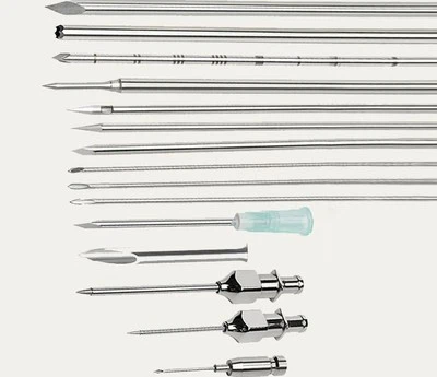
In 1980, Henning performed the first arthroscopic meniscus suture using a modified spinal needle and standard suture. Though technically crude, this marked the formal entryof meniscus treatment into the repair era. Over the following decade, a varietyof repair techniques emerged: inside-out suturing, outside-in suturing, bioabsorbable arrows, and meniscus staples.
Phase Four: Evidence-Based Medicine and Standardization (1990s–2010s) - From Experience to Evidence
Entering the 21st century, with the riseof evidence-based medicine, meniscus repair entered a standardized era. Sufficient clinical data allowed key questions to be answered:
Long-term outcomes: 10-year follow-up studies showed success rates around 85%, significantly lowering arthritis risk.
Key influencing factors: Vascular zone, tear pattern, and concurrent ACL reconstruction were most critical.
Technique comparison: In experienced hands, primary techniques yielded comparable results.
In 2005, the International Meniscus Repair Consensus Group published guidelines defining the "ideal candidate" for repair: young patient, acute tear (<8 weeks), vertical longitudinal pattern in red/red-white zone, length 1–4 cm, combined with ACL reconstruction. While strict, this profile yielded healing rates exceeding 90%.
Phase Five: The Era of Biological Augmentation (2010s–Present) - Beyond Mechanical Fixation
Over the last decade, the greatest advances have occurred not in mechanical technique, but in biological understanding. Research revealed that even "perfect" suturing results in fibrovascular scar tissue rather than native fibrocartilage, with mechanical properties recovering to only ~80% of normal.
This sparked the concept of "biological augmentation" - enhancing the local healing environment to improve repair quality.
Vascularity Enhancement: Rasping tear edges to create bleeding beds, converting avascular white zones into "pseudo-red zones," increasing healing rates by 20–30%.
Growth Factor Application: Platelet-rich plasma (PRP) and fibrin clots provide anabolic cytokines; meta-analyses show a 10–15% improvement in healing rates.
Stem Cell Therapy Exploration: Bone marrow-derived MSCs and adipose-derived stem cells are under preclinical investigation for their potential to differentiate into fibrochondrocytes.
Phase Six: Precision and Intelligence (Ongoing) - The Future of Personalized Repair
Current technological frontiers focus on intelligence and personalization.
Real-Time Navigation Systems: Electromagnetic or optical tracking of instrument tips, especially valuable in high-risk posterior horn repairs.
Mechanosensory Sutures: Monitoring postoperative tension changes to guide personalized rehabilitation.
3D Printing: Patient-specific guides manufactured from CT data to ensure precise entry angles and depths.
Historical Insights: A Spiraling Trajectory of Cognition
Reviewing this century-long evolution reveals a clear, spiraling trajectory:
Cycle One: From "Total Resection" (technical limitation) to "Cognitive Breakthrough" (recognition of functional importance).
Cycle Two: From "Partial Resection" (functional preservation) to "Repair Attempts" (deepening biological understanding).
Cycle Three: From "Simple Repair" to "Biological Augmentation" (integration of regenerative medicine).
Each cycle represents not just technological progress, but a philosophical shift. From viewing the meniscus as a dispensable remnant to recognizing it as a crucial guardian of long-term joint health, this transformation is rooted in decades of research, clinical practice, and long-term patient data.
Final Reflection
Perhaps the deepest lesson from the history of meniscus treatment is this: in medicine, a deep understandingof normal structure and function is the prerequisite for rational treatment. When we deem a structure "useless," we resort to the simplest, crudest solutions; only when we truly grasp its value do we invest the immense effort required to protect and restore it.
Today's meniscus repair surgery, with all its limitations, has preserved knee function and delayed the progression of arthritis for countless patients. This ongoing history will continue to witness how medical wisdom breaks through biological constraints to create better therapeutic possibilities.
If you would like, I can now compile all your translated sections - including this historical piece - into one comprehensive, journal-ready monograph with unified structure, references, and academic formatting.
Do you want me to proceed with that final integrated manuscript?