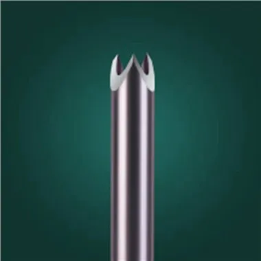
Sample Chamber
Sep 18, 2026
Pain Point - The Chamber That Crushes
The sample chamber and stylet notch are often confused, but they serve distinct functions. If the chamber volume is wrong, the core is short, crushed, or contaminated with blood. Pathology receives "core-like" material with no architectural integrity. The pain point is that many OEMs size the chamber based on inner diameter math alone, ignoring tissue mechanics. A chamber that is too long creates excessive vacuum, drawing blood into the specimen. A chamber that is too short yields inadequate length. A chamber with rough walls fragments the core during extraction. In prostate biopsies, where multiple cores are taken systematically from different zones, a crushed or bloody specimen can lead to inaccurate Gleason scoring, potentially resulting in either overtreatment (radical prostatectomy for a low-grade cancer) or undertreatment (active surveillance for a high-grade cancer). In breast biopsies, the sample chamber must preserve the architecture of microcalcifications, which are critical for diagnosing ductal carcinoma in situ (DCIS). If the chamber walls are too rough or the edge radius too large, the microcalcifications may be dislodged or the tissue crushed, leading to a false-negative result. For OEMs, the cost of a poorly designed sample chamber is high: pathologist complaints, reduced adoption by key opinion leaders, and the need for costly redesigns late in the development cycle. The sample chamber is not just a recess; it is a precision-engineered volume that must balance tissue capture, specimen integrity, and ease of extraction.
Principle - Controlled Dead Volume
The sample chamber is a controlled dead volume. Length × depth × wall clearance determines specimen quality. Laser-cut hypotube stylets allow engineers to tune chamber walls with radial cuts for tissue grip and interrupted spirals for stylet stiffness. The principle is to match chamber geometry to tissue elasticity: stiffer tissues require shorter, deeper chambers; softer tissues require longer, shallower chambers. The underlying biomechanics involve the interplay between tissue elasticity, interstitial pressure, and the mechanical properties of the chamber walls. When the stylet is inserted into the tissue, the surrounding tissue prolapses into the chamber due to the pressure differential. The rate and extent of prolapse depend on the chamber's volume and the tissue's compliance. Once the tissue is inside, the outer cannula fires forward, shearing the base of the core. The chamber walls must then hold the specimen securely during withdrawal but release it cleanly into the pathology cassette. Laser-cut hypotubes enable this functionality through precise geometric control. Radial cuts on the chamber walls can create micro-grips that prevent the specimen from slipping out prematurely, while interrupted spiral cuts on the stylet body provide the necessary stiffness to prevent buckling during insertion. The material choice - 316L for general use, 17-7PH for high stiffness, or Nitinol for flexible applications - further tailors the chamber's performance. The 0.012 mm kerf width ensures that the cuts are clean and the heat-affected zone minimal, preserving the fatigue life of the stylet.
Equipment Classification - Manufacturing the Chamber
(1) Fiber Laser Cutters - These machines cut the sample chamber profile with high precision, achieving the required length and depth tolerances (typically ±0.01 mm). The laser parameters are optimized for the specific material to minimize recast layer. (2) Notch Grinders - For chambers that require a ground floor rather than a laser-cut edge, CNC grinders provide the necessary edge radius control (typically <5 µm) to prevent tissue crushing. (3) Electrochemical Deburring (ECD) Systems - These remove any remaining burrs from the laser cutting process, ensuring a smooth chamber interior that will not fragment the specimen. (4) Volumetric Simulators - A test rig that uses a calibrated fluid or gel to measure the actual volume of the chamber, verifying that it matches the design intent. (5) Histology Scoring Panels - A group of pathologists who evaluate simulated biopsy cores for length, fragmentation, and architectural preservation. This is the ultimate validation of the chamber design.
Practical Guide - Designing the Sample Chamber
Step 1: Match chamber to gauge. For an 18 G needle, the core diameter is typically 1–1.5 mm; for 14 G, it is about 2 mm. The chamber depth should be 30–60% of the wall thickness, depending on tissue density. Step 2: Optimize chamber length. For most soft tissues, a length of 15–20 mm is sufficient. For firmer tissues like prostate, 10–15 mm may be adequate. Step 3: Specify surface finish. The chamber walls should be electropolished to Ra < 0.2 µm to prevent tissue adhesion. Step 4: Add retention features. Consider laser-cut radial grooves or micro-teeth on the chamber walls to help retain the specimen during withdrawal. Step 5: Validate with tissue phantom. Fire the needle into a calibrated gel and measure the extracted core's length and integrity. The core should be >80% of the chamber length and show minimal fragmentation. Step 6: Pathologist review. Have a pathologist evaluate the simulated cores to ensure they are suitable for diagnostic purposes.
Real-World Experience - Engineering for Architecture
A prostate CNB program was experiencing a high rate of bloody fragments, making Gleason scoring difficult. Investigation revealed that the sample chamber was too long (25 mm for an 18 G needle), creating excessive vacuum that drew blood into the specimen. Shortening the chamber to 18 mm and polishing the inner walls to Ra 0.15 µm resulted in clean, intact cores with minimal blood contamination. Pathologists reported "excellent architectural preservation, suitable for accurate grading." In another case, a breast biopsy device was producing cores with crushed margins. The chamber walls were too thin, and the laser-cut edge radius was too large (>10 µm). Switching to a thicker-wall tube and using a picosecond laser to reduce the edge radius to <5 µm eliminated the crushing artifact. These examples highlight the importance of treating the sample chamber as a precision component, not an afterthought.
Summary - Specimen Science
Sample chamber design is specimen science, not just machining. It determines whether pathology receives a diagnosis or a disappointment. A well-designed chamber captures the tissue gently, holds it securely, and releases it cleanly, enabling the pathologist to provide an accurate diagnosis. For manufacturers, investing in chamber optimization is an investment in clinical success.
Outlook - The Intelligent Chamber
Future sample chambers will be vacuum-assisted with adjustable volume, allowing the physician to adapt to tissue type in real time. Shape-memory chambers will change geometry during firing, expanding to capture more tissue or contracting to protect fragile specimens. Molecular-test-ready chambers will preserve RNA/DNA integrity for genomic profiling, bridging the gap between histopathology and molecular diagnostics. As personalized medicine advances, the sample chamber will become a critical link in the chain of diagnosis.