Precision Instruments: The Design Principles And Evolution Of Soft Tissue Biopsy Needles
Jun 16, 2026
The soft tissue biopsy needle, seemingly a simple medical device, is in fact one of the cornerstones of modern precision medicine. It is not a singular object but a sophisticated tissue acquisition system. Its design philosophy and evolutionary history profoundly reflect humanity's deepening understanding of disease and the relentless pursuit of minimally invasive technology.
Core Design: Balancing "Acquisition" and "Trauma"
The fundamental challenge in designing any biopsy needle lies in resolving a core contradiction: how to obtain sufficient, intact tissue samples for pathological diagnosis while minimizing patient trauma. This is manifested in several key parameters:
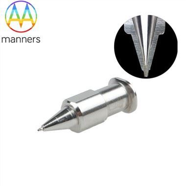
- Needle Tip Geometry: Early needle tips were merely simple beveled cutting edges. Modern biopsy needles have evolved into complex geometries, such as the sharp Trocar tip designed for penetrating dense tissues, or Franseen-style and Core-style tips featuring grooves or barbs that effectively capture and retain the tissue core during withdrawal, preventing sample loss. This is critical for achieving "successful sampling on the first attempt."
- Cutting Mechanism: The evolution spans from manual rotation to semi-automatic and fully automatic spring-loaded mechanisms. Manual needles rely heavily on operator feel, resulting in inconsistent sample quality. Semi-automatic needles utilize spring mechanisms to achieve rapid cutting, reducing human tremor. Fully automatic firing needles (such as the Bard® Mission®) deliver millimeter-level precision in throw distance and speed, significantly increasing the success rate for deep-seated small lesions.
- Guidance and Localization: Initial blind punctures relied on anatomical landmarks, posing high risks. Today, image guidance using Ultrasound (US), Computed Tomography (CT), and Magnetic Resonance Imaging (MRI) has become the standard. Specialized echogenic markers on the needle shaft enhance visibility under ultrasound, while CT-compatible carbon fiber or titanium alloy shafts prevent artifact interference. Advanced biopsy guns integrate coaxial cannula systems, allowing multiple samples to be taken without repeated punctures, drastically reducing complications such as pneumothorax and hemorrhage.
Empowerment Through Materials Science
As referenced in your provided materials, material selection defines the performance boundaries of biopsy needles.
- Stainless Steel (304/316L): Remains the workhorse. Its high rigidity ensures directional stability during penetration, while its corrosion resistance and low cost make it ideal for disposable products. However, brittleness becomes a challenge for extremely fine gauges (e.g., 25G and above).
- Titanium Alloy (Ti-6Al-4V): Its revolutionary advantage lies in MRI compatibility. For interventional procedures requiring operation within strong magnetic fields, titanium alloy needles are the only safe choice. Furthermore, their superior strength-to-weight ratio allows for thinner walls while maintaining rigidity, thereby increasing the inner diameter to procure thicker tissue cores.
- Medical-Grade Polymers (e.g., PEEK, Polycarbonate): Primarily used for handles, coaxial cannulas, and hubs. They provide insulation, lightweight handling, ergonomic grip, and cost efficiency. Novel biodegradable polymers are currently under investigation for temporary implantable micro-biopsy devices.
Evolutionary Trajectory: From "Acquiring" to "Targeting" to "Optimizing"
- 1950s–1970s: Represented by manual split-needle designs like the Vim-Silverman needle, characterized by significant trauma and low success rates.
- 1980s–1990s: The advent of the Tru-Cut™ semi-automatic biopsy needle was a milestone. Featuring an integrated tissue notch and cutting cannula, it standardized sampling. Subsequently, fully automatic spring-loaded biopsy guns (e.g., Biopty® Gun) simplified the procedure to a trigger pull, significantly flattening the learning curve for physicians.
- 21st Century to Present: Development focuses on:
- Miniaturization: Developing finer needles (e.g., 25G) for high-risk areas such as the brainstem and pancreas.
- Intelligence: Integrating pressure sensors or optical probes to provide real-time feedback on tissue stiffness or spectral characteristics, assisting in verifying target engagement.
- Targeted Therapy Integration: Combining with navigation systems and robotic-assisted platforms to achieve stereotactic biopsies with sub-millimeter precision.
In summary, the design of the soft tissue biopsy needle represents a continuous quest for the optimal equilibrium between "minimally invasive" techniques and "precision." Every material innovation and every mechanical optimization pushes the boundaries of pathological diagnosis forward, enabling more patients to benefit from early and accurate detection.