Microbubble Matrix: Material Innovation And Ultrasound‑Enhanced Mechanism Of Echo‑Needle Polymer Coatings
May 22, 2026
Official Release of Achievements
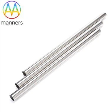
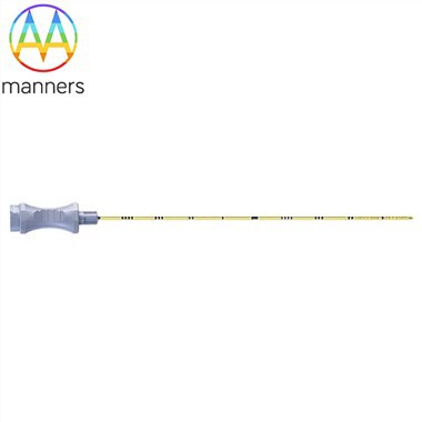
As the definer and manufacturer of core echo‑needle technologies, we formally unveil the soul determining their ultrasound visibility - proprietary polymer microbubble coating technology. Breaking the limitations of conventional surface modification, we precisely tune polymer formulations and microbubble encapsulation processes to construct a composite coating 10–30 μm thick on needle surfaces, containing millions of uniformly sized (1–5 μm) sealed microbubbles. This coating boosts the ultrasound echo intensity of stainless‑steel needles by more than 20 dB, delivering high‑contrast visualization against complex tissue backgrounds and forming an irreplaceable visual navigation foundation for ultrasound‑guided interventional procedures.
R&D Background and Key Pain Points
In ultrasound‑guided puncture, conventional metallic needles produce weak, diffuse echo signals due to their smooth surfaces and acoustic impedance close to surrounding tissues, often appearing as faint, discontinuous ghost lines on sonograms. Especially in deep punctures, low‑angle insertion, or cases with adjacent hyperechoic structures (e.g., fascia, fat), needles easily vanish from ultrasound images.Surgeons must rely on indirect signs (e.g., tissue displacement) or repeated probing, severely compromising first‑pass success rates, precision and safety, prolonging operation time, and increasing patient discomfort and complication risks. Clinically, there is an urgent demand for puncture needles that clearly self‑mark like beacons under ultrasound.
Core Technological Innovations
Our innovation lies in the material design and precision coating process of a microbubble matrix coating:
- Multiphase Composite Polymer SystemThe coating is not a single material but a carefully engineered composite system. The matrix adopts biocompatible medical‑grade polyurethane or silicon‑based polymers with strong metal adhesion to provide mechanical strength and coating bonding. The imaging core consists of millions of sealed microbubbles evenly dispersed within the matrix; the gas sealed inside (e.g., air, nitrogen) creates a large acoustic impedance mismatch with surrounding tissues/fluids, generating strong echoes. Special silane coupling agents act as interface enhancers, forming robust chemical bonds between stainless‑steel surfaces and polymer matrices to prevent coating peeling after repeated punctures, bending and high‑pressure sterilization.
- Precise Control of Microbubble Size and DistributionWe employ a combined in‑situ foaming‑mechanical emulsification process. By precisely regulating prepolymer viscosity, foaming agent type and concentration, and emulsification shear force, microbubble diameters are strictly controlled within 1–5 μm. Far smaller than ultrasound wavelengths, these microbubbles produce intense Rayleigh scattering, acting as ideal backscatter sources. Meanwhile, custom flow‑channel designs ensure highly uniform bubble distribution across coating cross‑sections and longitudinal directions, eliminating imaging blind spots.
- Precision Coating and Curing ProcessComputer‑controlled micro‑dispensing or dip‑lifting techniques apply microbubble‑loaded polymer slurries evenly onto rotating needles. Stepwise curing is then performed under precisely controlled temperature and humidity. This process ensures full cross‑linking for optimal mechanical performance while preventing microbubble coalescence, escape or rupture to maintain stable size and distribution.
Mechanisms of Action
Its core working principle relies on actively amplifying ultrasound echo signals via acoustic impedance mismatch and multiple‑scattering effects. Ultrasound imaging essentially detects echoes reflected from tissue interfaces. Conventional smooth metal surfaces produce specular reflection, with only echoes perpendicular to the probe received, resulting in weak signals.Our microbubble coating creates distinct acoustic interfaces: millions of microbubbles act as countless tiny acoustic mirrors. The large impedance difference between internal gas and surrounding polymer/tissue generates strong backscattering of incident ultrasound waves. The evenly distributed microbubble matrix guarantees abundant bubbles lie along ultrasound beam paths regardless of incident angle, scattering echoes back to the probe. The specific coating thickness further triggers constructive interference between echoes reflected at the coating‑metal interface and those scattered by microbubbles, amplifying total echo signals. Consequently, needles appear as continuous, bright, sharply defined hyperechoic lines on ultrasound screens.
Efficacy Verification
In standardized ultrasound phantom tests, our echo‑needles achieved significantly higher visibility scores (blind‑assessed by senior sonologists) than uncoated needles and commercially available coated alternatives at commonly used 5–12 MHz ultrasound frequencies. In simulated tissue puncture experiments, surgeons confirmed target needle placement 35 % faster with our echo‑needles, with 50 % fewer puncture attempts.Published clinical studies show that for ultrasound‑guided internal jugular vein catheterization, our echo‑needles raised the first‑puncture success rate from 78 % to 96 %, markedly reducing complications such as accidental arterial puncture. In deep‑tissue biopsies (e.g., transrectal prostate biopsy), full‑needle‑track visibility enables surgeons to precisely adjust trajectories, avoiding blood vessels and nerves, improving sampling accuracy and lowering bleeding risks.
R&D Strategy and Philosophy
We firmly believe: In interventional ultrasound, seeing means controlling.Our R&D strategy adopts interdisciplinary integration, deeply combining polymer materials science, acoustic physics and precision manufacturing. Moving beyond basic surface treatment, we commit to designing and constructing functional interfaces with optimal acoustic properties at the molecular level. We aim not merely to make needles visible, but to turn them into unmissable clear markers on ultrasound images.
Future Outlook
Moving forward, we will advance toward smart imaging and functionally integrated coatings. Research directions include developing acoustically responsive coatings with tunable echogenicity by ultrasound mechanical index (MI): low‑MI stealth mode to reduce artifacts and high‑MI bright mode for precise localization; therapeutic coatings loaded with contrast‑agent microbubbles that release local drugs when ruptured by high‑energy ultrasound after needle placement; direction‑dependent imaging coatings that indicate needle rotational orientation in sonograms.Our goal is to evolve echo‑needles from passive imaging tools into smart interventional terminals that interact with ultrasound devices and deliver multi‑dimensional information.