In-Depth Analysis Of Acoustic Scattering, Micro-Etching & Polymer Coating Visualization Mechanisms
Jul 05, 2026
https://www.nature.com/articles/s41598-024-72620-8
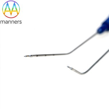
That echogenic needles appear as bright lines rather than "invisible" on ultrasound stems from deliberate acoustic interface engineering and material modification. To understand the principle, one must first understand acoustic impedance mismatch and the limitation of specular reflection: the impedance difference between metal and soft tissue is enormous-theoretically producing strong reflection-but a smooth cylindrical needle surface only returns a detectable echo when the ultrasound beam is nearly perpendicular. Once the insertion angle steepens (common in out-of-plane puncture), the reflected wave deflects away from the probe aperture per the law of equal incident/reflection angles, causing partial or complete loss of the needle image-the clinically familiar "needle drop-out."
Mechanism 1: Mechanical Micro-Etching / Micro-Dimpling (Micro-Dimpling / Laser Etching)
Manufacturers use precision CNC or laser processing to create regularly arranged micron-scale pits, grooves, or honeycomb arrays on the needle tip and predetermined visualization zone of the shaft. These microscopic geometric features break up the continuous mirror surface, causing incident ultrasound to diffract and scatter at multiple points, with part of the energy radiating in all directions-including back to the probe. Well-designed micro-etching maintains clear echoes at large deviation angles of 30°–70°, and doubly reinforced tip etching produces a bright dot facilitating depth determination. The slight increase in roughness is a theoretical drawback; isolated reports mention minimally increased tissue drag, though clinical impact is generally negligible.
Mechanism 2: Polymer Coating with Microbubbles / Ceramic Microspheres (Polymer Echogenic Coating)
Medical polyurethane or silicone-based polymers are blended with closed microbubbles (micro-air-bubbles), hollow glass microspheres, or high-impedance ceramic particles, and uniformly coated on the needle shaft (usually the visualization segment). Countless micro-interfaces within the coating create extensive acoustic impedance discontinuities; incident waves generate intense backscatter, rendering the shaft as a continuous bright line on ultrasound. Representative commercial technologies include Cook Medical's EchoTip® series and Encapson's Sono-Coat™. The coating method does not compromise the substrate, can be applied to needles with side ports/sample notches, and microbubble size/density can be optimized for target depth-smaller, denser bubbles for shallow targets (fine bright line), larger bubbles for deeper targets (enhanced echo, possibly with mild posterior shadowing).
Mechanism 3: Bulk Material / Composite Structure Modification (Less Common)
Research-stage approaches include porous metal sintering or composite embedding to give the needle body intrinsic scattering ability, but current mass production centers on etched + coated combinations.
Key Variables Affecting Visibility:
- Insertion Angle: Smaller angles (near-parallel to beam) allow some visibility even with plain needles; steeper angles depend heavily on echogenic enhancement-micro-etching shows greater advantage at large angles.
- Probe Frequency: High-frequency linear array (7–15 MHz) for superficial nerve blocks/vascular access; low-frequency convex array (2–5 MHz) for deep organs requires stronger scattering coatings.
- Needle Gauge: Coarse needles (14–18G) have larger scattering cross-sections and are easier to see; fine needles (22–25G) critically depend on high-quality echogenic treatment.
- Tissue Background: Fat-infiltrated liver or retroperitoneal hyperechoic backgrounds reduce contrast, demanding higher coating reflectivity.
- Clinical Tip: Even with echogenic needles, keeping the shaft within the ultrasound scan plane (in-plane technique) remains best practice; in out-of-plane technique, rely on the reinforced tip echo dot for depth judgment. Newer probes with "Needle Guidance / Beam Steering" functionality can further compensate.
In conclusion, echogenic needle visualization is not "magic" but controlled acoustic scattering engineering-micro-dimples decompose the mirror into diffuse sources, microbubble polymer coatings provide abundant backscattering interfaces, and either alone or in combination they make the otherwise "disappearing needle" stably glow on the ultrasound screen, furnishing interventionalists with real-time spatial localization cues.