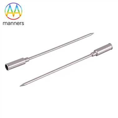
Huber Point Needles: Choosing 8G To 18G
Nov 21, 2026
Meta description: How to select Huber point needles from 8G to 18G for marrow biopsy, aspiration and port access, including core size and patient factors.
Gauge is the first specification most buyers discuss and the last one most understand. Two needles labelled 11G can differ in wall thickness, inner bore, tip geometry and effective sample volume, and those differences decide whether a biopsy yields an interpretable core or a fragment. Choosing within the 8G to 18G range used by Huber Point Needles is therefore a clinical decision expressed as a number, not a purchasing convenience.
The Cost of Choosing by Habit
Units develop habits. One department standardises on 11G because it was the gauge in the original kit, another uses 8G for every marrow biopsy because larger cores feel more informative. Neither habit is wrong in itself, but both become costly when applied without regard to the patient. A large-bore needle in a thin, osteoporotic patient increases penetration difficulty and post-procedure discomfort without adding diagnostic value.
The opposite error is subtler. A small gauge that shortens the procedure may yield a core too short or too fragmented for architecture assessment, forcing a repeat. The repeat costs more than the larger needle would have, and it exposes the patient twice. Between these poles sits a selection logic based on what the specimen must answer, not on what the shelf happens to hold.
What Gauge Actually Controls
Outer diameter sets the track and the force required. An 8G needle near four millimetres needs a firm, well-controlled push and a secure landmark; an 11G near three millimetres is easier to place and still yields a substantial core. A 13G near 2.4 millimetres balances sample volume against comfort, while 14G and finer are generally aspiration instruments rather than histological tools.
Inner diameter sets the specimen. Wall thickness consumes part of the nominal size, so a thin-walled 11G can deliver a core closer to a thick-walled 8G than the gauge labels suggest. For biopsy work the inner bore must also be smooth and consistent along its length; a slight taper from swaging or drawing will grip the core and cause it to break during extraction.
Length and tip complete the picture. Marrow needles are typically short enough to reach the iliac crest without excessive leverage, while port needles are supplied in lengths matched to tissue depth. A tip optimised for one application does not automatically suit the other, even at the same gauge, because cortical bone and silicone septum present entirely different resistance profiles.
Wall thickness is the hidden variable that purchasing teams most often miss. Two 11G cannulas can differ by several hundredths of a millimetre in wall, which changes both the core diameter and the force needed to push the needle through cortex. The specification should therefore state outer diameter, inner diameter and concentricity, not gauge alone. A supplier who can quote all three is usually the one who controls the drawing rather than assembling from bought-in tube.
A Working Classification
For marrow work, 8G suits large-volume histology where cellularity and architecture must be assessed in a single pass, and where the cortex is accessible and the patient stable. It demands more force and a confident technique. The 11G is the general-purpose compromise: adequate core, manageable force, wide availability in kit form. The 13G serves narrower anatomy, paediatric cases and situations where a smaller track is preferred.
For aspiration, 14G to 16G allow rapid sampling with less trauma, while 18G supports fine aspiration and some paediatric applications. In port access, 19G to 22G dominate because the septum tolerates small, non-coring punctures better and dwell comfort improves with a finer needle. The same number therefore carries different meaning in different product families, which is why catalogue selection should always state the application alongside the gauge.
Kits add a further layer. A gauge that is optimal in a standalone needle may be awkward inside a kit whose removal guide or safety helmet is sized for another bore. Compatibility across the whole assembly, not just the needle, determines whether the larger core can actually be extracted without damage.
Practical Selection Guide
Begin with the clinical question. If the pathologist needs architecture, cellularity and reticulin grading from one core, choose the largest gauge the patient's anatomy and cortex will tolerate comfortably. If the question is mainly cellularity or staging with limited material, 11G or 13G is usually sufficient and faster.
Then assess the patient. Body habitus affects landmark depth; bone quality affects penetration force; anticoagulation and thrombocytopenia affect bleeding risk and therefore track size. Age and cooperation influence how much force can be applied safely. A gauge decision that ignores these factors will be revised in the procedure room, usually at the worst moment.
Next, check the assembly. Confirm that the stylet fits without a gap, the removal guide passes the inner bore freely, and the safety helmet locks without requiring force. Verify that the Luer lock cone accepts the syringes in use. Finally, record the gauge on the request form and in the report so that subsequent procedures and audits can compare like with like.
Field Experience
Departments that switched from routine 8G to routine 11G often report fewer repeat passes, shorter procedures and similar diagnostic yield, because the smaller needle is easier to place accurately and produces less fragmentation. The gain comes from precision rather than from sample volume. Where 8G remains essential is in hypocellular marrow and in cases where a long, intact core is the only way to assess distribution.
Purchasing experience points in the same direction. Over-specifying gauge drives cost without proportional benefit, while under-specifying length creates waste through failed access. The most useful supplier conversation is not about price per unit but about which gauge, length and tip combination the supplier can hold consistently, and what evidence exists that the inner bore is uniform lot to lot.
Inventory practice matters as much as clinical logic. A department that stocks two gauges, two lengths and one kit configuration can cover most adult and paediatric needs, and it can train staff to recognise each item quickly. Long catalogues of near-identical sizes invite selection errors, and errors in gauge are expensive because they are discovered only after the needle is in the bone.
Age-related bone change adds a further dimension. Elderly patients often have a thin, brittle cortex over a fatty marrow space, so a large needle may penetrate easily but yield a core that fragments during processing. Younger patients may have denser bone that resists entry and rewards a sharper, more robust tip. Neither group is served by a single default, and a protocol that records bone quality alongside gauge builds useful experience for the next case.
Summary
Gauge selection in Huber Point Needles is a trade between specimen requirement and procedural burden. Outer diameter governs force and track, inner diameter governs core quality, and kit compatibility governs whether the sample survives extraction. A reasoned choice among 8G, 11G, 13G and the finer aspiration sizes will outperform a habitual one on both diagnostic yield and patient experience.
Outlook
Colour coding and clear laser marking of gauge, length and lot are becoming expected rather than optional, because they reduce selection errors in storage rooms and procedure trolleys. Kits configured around a single gauge with interchangeable guides simplify inventory and remove the temptation to mix components.
Selection may also become more evidence-based. Registries that record gauge, needle lot and specimen adequacy could show which combinations work best in which patient groups, turning a matter of local habit into a measurable choice.
Longer term, imaging and depth measurement may allow gauge and length to be chosen from patient data rather than from averages. Until that arrives, the practical route is a documented selection protocol, a small validated product range and a supplier able to demonstrate bore consistency. Those three elements solve most of the problems that gauge alone cannot. They also give a department a defensible answer when a clinician asks why one needle was chosen over another, which matters more than a catalogue number when practice is audited and when stock levels are reviewed.